Mar 27, 2023
How cell mechanics influences everything
Posted by Jose Ruben Rodriguez Fuentes in categories: bioengineering, biotech/medical, chemistry
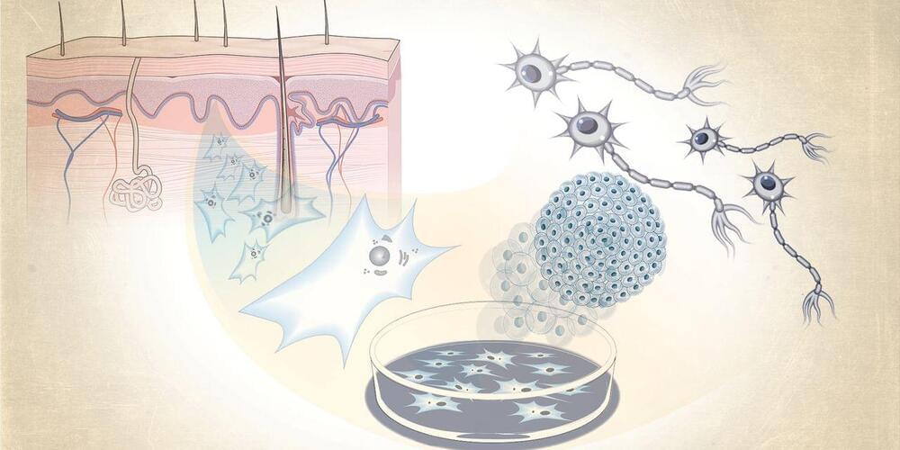
“People study cells in the context of their biology and biochemistry, but cells are also simply physical objects you can touch and feel,” Guo says. “Just like when we construct a house, we use different materials to have different properties. A similar rule must apply to cells when forming tissues and organs. But really, not much is known about this process.”
His work in cell mechanics led him to MIT, where he recently received tenure and is the Class of ’54 Career Development Associate Professor in the Department of Mechanical Engineering.
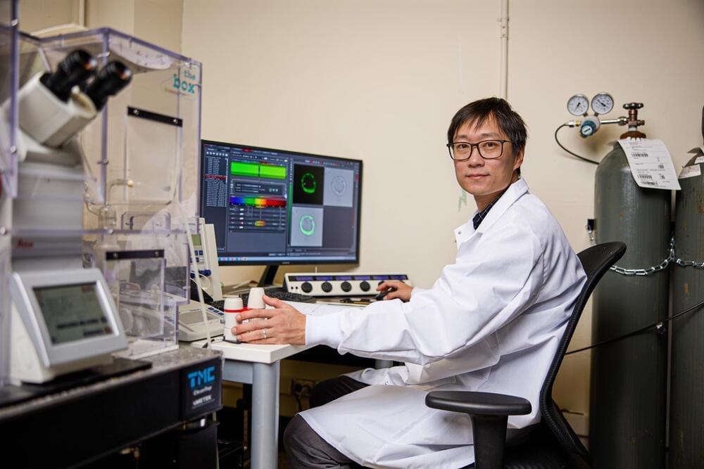
At MIT, Guo and his students are developing tools to carefully poke and prod cells, and observe how their physical form influences the growth of a tissue, organism, or disease such as cancer. His research bridges multiple fields, including cell biology, physics, and mechanical engineering, and he is working to apply the insights from cell mechanics to engineer materials for biomedical applications, such as therapies to halt the growth and spread of diseased and cancerous cells.
