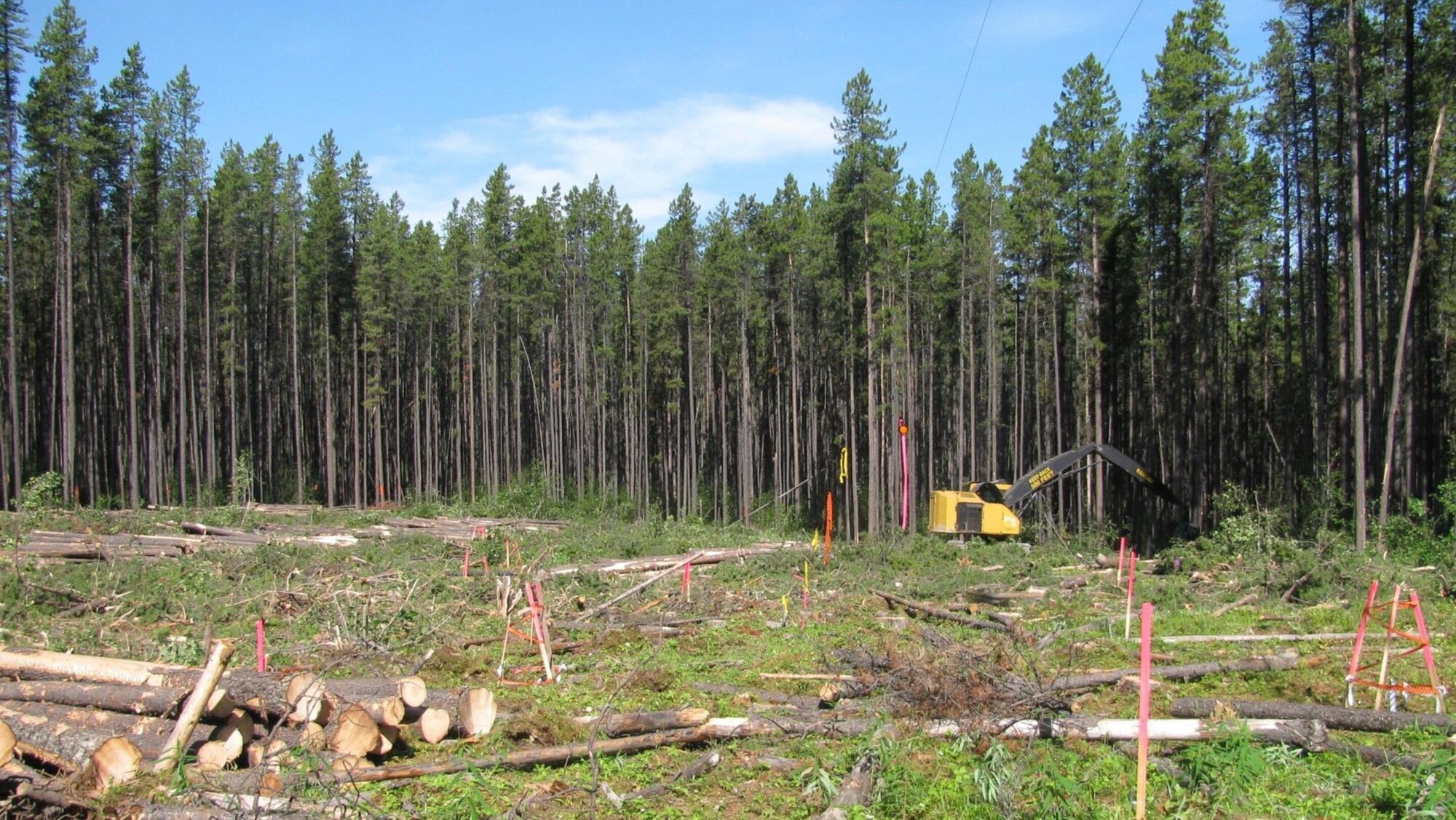
Toggle light / dark theme

Cell death’s ‘beautiful’ rings!

Over the past several decades, researchers have identified the genes and proteins in plants that initiate the cellular self-destruct sequence. During that time, they also found shared elements of this “resistome” at work in mammalian.

Plant nucleotide-binding leucine-rich repeat (NLR) immune receptors detect pathogen effectors and activate immunity. Coiled-coil NLRs (CNLs) form resistosomes as Ca2+-permeable channels in the plasma membrane (PM). However, the mechanism by which resistosomes activate cell death remains unclear.

The ring, which resembles a wreath or a necklace, the author said, is a combination of proteins that bind to a cell membrane and six channels that orient themselves to run through the membrane. The team made this discovery working with Arabidopsis and Nicotiana bethamaian, popular plant model systems, and a high resolution total internal reflection fluorescence microscope.

The authors show that the CNL SUPPRESSOR OF mkk1 mkk2 2 (SUMM2), unlike canonical CNLs that use a MADA motif to penetrate the PM, tethers to the PM through N-myristoylation, a common feature among many CNLs.

PM targeting via N-myristoylation is essential for SUMM2-induced cell death. Upon activation, SUMM2 promotes the association of the lipase-like proteins ENHANCED DISEASE SUSCEPTIBILITY 1 (EDS1) and PHYTOALEXIN DEFICIENT 4 (PAD4) with the helper NLR-ACTIVATED DISEASE RESISTANCE 1-LIKE 1 (ADR1-L1).

Active SUMM2 induces the clustering of multiple ADR1-L1 resistosomes into a ring-like assembly colocalized with the EDS1–PAD4 complex, and the EDS1–PAD4–ADR1 module is essential for SUMM2-activated cell death.

The finding invites new questions about what exactly the rings do and how they do it. The team’s current hypothesis is that the rings enable communication with nearby cells, sending inflammation signals that can help initiate cell death in a targeted way. ScienceMission sciencenewshighlights.

Bacteria that generate electricity: How a shellfish-based gel could monitor wastewater and food

Microbial bioelectronic sensors use living bacteria that can create an electrical signal in response to the presence of a target substance, or analyte. These types of sensors offer many advantages over other types of biosensors based on proteins and enzymes: The bacteria can perform multiple functions, survive in a variety of environments and even grow and regenerate for potential long-term use.

However, building devices using living bacteria poses several challenges. The mediators some bacteria use to send and receive electrons, creating the electric signal, can be swept away from the sensor by liquid environments researchers would want to monitor, like wastewater. Some mediators are toxic to humans or the environment. Rice University researcher Rafael Verduzco developed a safe bioelectronic sensor that allows for effective electronic communication even in liquid environments. The study was recently published in the journal Advanced Materials.

“This system uses a naturally occurring polymer chitosan, which is found in the hard outer shells of crustaceans. In our system, the chitosan also acts kind of like a shell to keep the bacteria from escaping. It is also modified to have anchor points the mediators can attach to, which are critical to transport electrons,” said Verduzco, corresponding author on the paper and the A.J. Hartsook Professor of Chemical and Biomolecular Engineering. “This material provides a flexible way to encapsulate the bacteria and enhance electronic signals. Since it’s based on a low-cost and renewable polymer, we think it has great potential for real-world applications.”

Enhancing gut-brain communication reversed cognitive decline, improved memory formation in aging mice

The sight of a delectable plate of lasagna or the aroma of a holiday ham are sure to get hungry bellies rumbling in anticipation of a feast to come. But although we’ve all experienced the sensation of “eating” with our eyes and noses before food meets mouth, much less is known about the information superhighway, known as the vagus nerve, that sends signals in the opposite direction — from your gut straight to your brain.

These signals relay more than just what you’ve eaten and when you are full. A new study in mice from researchers at Stanford Medicine and the Palo Alto, California-based Arc Institute has identified a critical link between the bacteria that live in your gut and the cognitive decline that often occurs with aging.

“Although memory loss is common with age, it affects people differently and at different ages,” said Christoph Thaiss, PhD, assistant professor of pathology. “We wanted to understand why some very old people remain cognitively sharp while other people see significant declines beginning in their 50s or 60s. What we learned is that the timeline of memory decline is not hardwired; it’s actively modulated in the body, and the gastrointestinal tract is a critical regulator of this process.”


By Krista Conger

Aging causes changes in gut bacteria in mice, which hampers communication between the intestines and the brain. Restoring this connection helped old mice form memories as well as young animals.

Pollen-replacing feed strengthens honey bee colonies, long-term study confirms

A man-made food source provided honey bees a nutritious diet at a commercial scale over the course of two winter seasons, according to a new study led by Washington State University researchers. The study, published in the journal Insects, looked at the new feed as used by five commercial beekeepers in California and Idaho from fall 2022 to spring 2024. This study is a follow-up to an initial paper describing the bee feed.

The nutritionally complete feed, which resembles an oversized, very thin granola bar, was developed by APIX Biosciences, a biotech company based in Belgium with a U.S. subsidiary. The company worked with WSU’s Honey Bee Program to test the nutritional supplement.

“The first paper was a trial during the spring and summer pollination season to make sure the feed worked in real-world field conditions,” said Brandon Hopkins, WSU’s P.F. Thurber Endowed Distinguished Professor of Pollinator Ecology and a corresponding author on the paper. “This study happened during the other half of the year when beekeepers tend to see the biggest losses and depend the most on supplemental feeding. It was also done on a significantly larger scale than our previous study.”

Lunar regolith simulant used to grow chickpeas

Dr. Sara Santos: “The research is about understanding the viability of growing crops on the moon. How do we transform this regolith into soil? What kinds of natural mechanisms can cause this conversion?” [ https://www.labroots.com/trending/space/30294/lunar-regolith…hickpeas-2](https://www.labroots.com/trending/space/30294/lunar-regolith…hickpeas-2)


How will astronauts grow food during long-term missions to the Moon? This is what a recent study published in Scientific Reports hopes to address as a team of scientists investigated the prospect of growing food on the Moon. This study has the potential to help scientists, mission planners, engineers, and astronauts develop new methods for growing food on the Moon, which could help advance such techniques when humans go to Mars.

For the study, the researchers grew chickpeas using simulated lunar regolith (often mistakenly called “soil”) and fungi, with the latter being used to test plant stress levels, decrease toxins, and enhance the mixture of regolith simulant and fungi. The team tested a variety of mixtures, including 25 to 100 percent regolith simulant and with and without the fungi. The goal of the study was to ascertain the plausibility of growing food on the Moon under climate-controlled conditions using lunar regolith and Earth-based products. In the end, the researchers found that the most promising mixture was 75 percent regolith simulant with fungi.

Electron microscopy maps protein landscapes that drive photosynthesis

Research led by scientists at Washington State University has revealed insights on how plants form a microscopic landscape of proteins crucial to photosynthesis, the basis of Earth’s food and energy chain. The discovery provides a new view of the molecular engine that converts sunlight into bioenergy and could enable future fine-tuning of crops for higher yields and other useful traits.

Colleagues at WSU, the University of Texas at Austin, and the Weizmann Institute of Science in Israel used a novel, technology-powered approach to peer inside plant leaf cells and visualize the landscape of the photosynthetic membrane—the ribbon-like structure where plants harvest sunlight. The findings were recently published in the journal Science Advances.

“These membranes are highly efficient biological solar cells,” said the study’s principal investigator and corresponding author, Helmut Kirchhoff. “They convert sunlight energy into chemical energy that fuels not only the plant’s metabolism but that of most life on Earth.”

Light-guided ‘optovolution’ evolves proteins that switch states on schedule

EPFL researchers have developed a light-based method that can produce proteins that switch states, respond to signals, and even compute, using light and the cell cycle.

Evolution is biology’s powerful method of engineering. It works by generating many variants of DNA, RNA, and proteins inside cells and letting nature “select” the organism that performs best. Early farmers started taking advantage of evolution by interfering with natural selection and letting only the most productive livestock and crops mate.

In laboratories, researchers have developed methods for directed evolution of proteins, especially enzymes and antibodies, that are used in household detergents, medicine, and industry.

Cannabis Extracts Significantly Reduce Myofascial Pain

“These findings indicate the clinical potential of cannabinoids as a promising therapeutic alternative for managing TMD…” [ https://www.labroots.com/trending/cannabis-sciences/30277/ca…ial-pain-2](https://www.labroots.com/trending/cannabis-sciences/30277/ca…ial-pain-2)


Temporomandibular Disorder (TMD) consists of a myriad of conditions causing jaw pain and dysfunction and the muscles controlling jaw movement. One type of pain is myofascial pain, which is associated with deep, aching muscle pain around the jaw, often resulting in later neck and shoulder pain. Traditional treatments include self-care like eating soft foods and over-the-counter anti-inflammatories. But how can Delta-9-Tetrahydrocannabinol (THC) and Cannabidiol (CBD) be used to relieve myofascial pain in patients suffering from TMD?

Now, a team of researchers from Brazil might be one step closer to better understanding the role of cannabis in treating myofascial pain. For their study, which was recently published in the journal Clinics, the team investigated how a combination of THC and CBD drug therapy could be used to not only decrease myofascial pain while also enabling TMD patients to regain jaw function. Over a 90-day period, 20 adults suffering from myofascial pain due to TMD were given gradual increases of THC and CBD starting from 2mg and eventually 10mg near the end of the trial.

The goal of the study was to ascertain the role of THC and CBD on treating myofascial pain and improving jaw movement and function. In the end, the researchers found that not only did the participants report an approximate 90 percent reduction in pain, but they were able to open their jaws approximately 4 mm wider than before the THC/CBD treatment.

Trapping light on thermal photodetectors shatters speed records

Electrical engineers at Duke University have demonstrated the fastest pyroelectric photodetector to date, which works by absorbing heat generated by incoming light. Capable of capturing light from the entire electromagnetic spectrum, the ultrathin device requires no external power, operates at room temperature and can be readily integrated into on-chip applications.

The advance could form the basis of a new class of multispectral cameras capable of impacting a wide range of fields such as skin cancer detection, food safety inspection and large-scale agriculture.

The results appear in Advanced Functional Materials.

Frontiers: Dietary restriction (DR)

Defined as reduced caloric intake or selective limitation of specific nutrients without malnutrition, is one of the most robust interventions known to extend lifespan and healthspan across species. Studies from yeast to mammals demonstrate that DR elicits conserved genetic, transcriptional, and epigenetic programs that promote cellular maintenance and stress resistance. At the molecular level, DR engages evolutionarily conserved nutrient-sensing pathways, including insulin/IGF-1 signaling (IIS), the mechanistic target of rapamycin (mTOR), AMP-activated protein kinase (AMPK), and NAD+-dependent sirtuins, which converge on key transcription factors (TFs) and transcriptional coactivators (TCs) to coordinate metabolic and longevity-associated gene expression. Downstream, these pathways enhance autophagy and proteostasis, remodel mitochondrial function and redox balance, reshape immune and inflammatory networks, and induce epigenetic and transcriptional reprogramming. Recent work further highlights amino acid–specific sensing mechanisms, endocrine mediators such as fibroblast growth factor 21 (FGF21), the gut microbiome, circadian regulators, and nuclear pore–associated transcriptional plasticity as integral components of DR responses. Importantly, the physiological outcomes of DR are context dependent and influenced by genetic background, sex, age at intervention, and the type and duration of restriction. In this review, we summarize current knowledge on the genetic and molecular architecture underlying DR-induced longevity and health benefits across species, discuss implications for aging-related diseases, and outline future directions toward precision nutrition and safe translational strategies.

Aging is characterized by a progressive decline in physiological integrity, reduced stress resilience, and increased susceptibility to chronic diseases (Lopez-Otin et al., 2023). Among numerous genetic, pharmacological, and lifestyle interventions examined over the past decades, dietary restriction (DR) remains the most robust and evolutionarily conserved strategy for extending lifespan and improving healthspan. Originally described in rodents nearly a century ago, the beneficial effects of reduced nutrient intake have since been validated in a wide range of organisms, including yeast, nematodes, flies, and mammals (Wu et al., 2022). While often used interchangeably, it is critical to distinguish between different nutritional interventions to avoid conceptual overlap. Caloric restriction (CR) typically refers to a chronic reduction in total calorie intake (usually 20%–40%) without malnutrition.

/* */