“The Commission considers that at this stage it cannot propose a legal obligation to keep videogames playable after they stop being provided commercially. This is due, also, to existing intellectual property rights. Under EU copyright law, rights holders enjoy exclusive rights over their creations.”
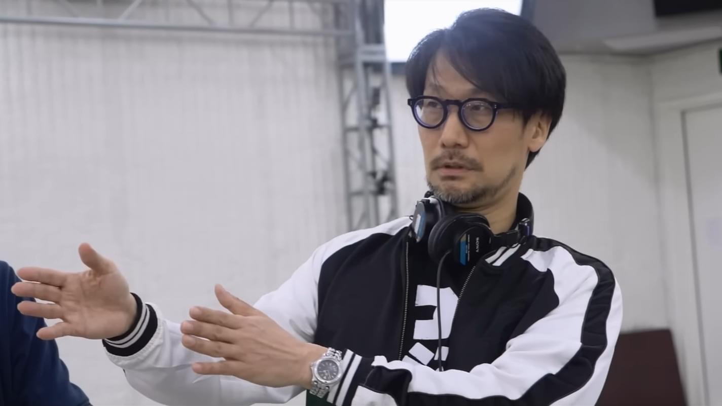
Players are (quite rightly) worried that without physical media their beloved games, or any kind of art, can be ripped away from them at a moment’s notice. “We will not be able to freely access the movies, books, and music that we have loved,” Kojima adds. “I would be a have-not. That’s what I’m afraid of. This is not greed.”