Toggle light / dark theme

Single-step 8-9x expansion reveals nanoscale centrioles without electron microscopy

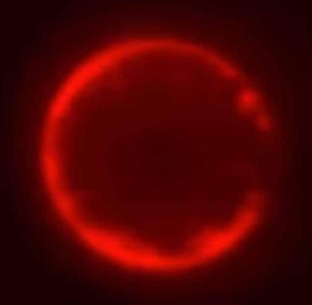
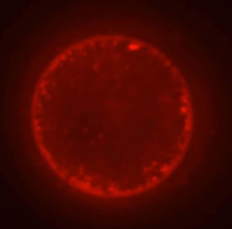
In a study published in ACS Nano, researchers from National Taiwan University report a new expansion microscopy strategy termed high-fold homogeneous expansion microscopy (hiHomoExM), capable of achieving approximately 8–9× isotropic expansion in a single expansion step while preserving delicate ultrastructural organization.

Expansion microscopy works by embedding biological samples within a swellable polymer hydrogel. Following chemical processing, the hydrogel expands uniformly in water, physically separating biomolecules and effectively increasing the spatial resolution achievable by conventional light microscopes.

“To achieve nanoscale imaging faithfully, both high expansion and homogeneous specimen preservation are essential,” explains the research team. “Nonuniform expansion can distort ultrastructural information and limit biological interpretation.”

MIT researchers use AI to uncover atomic defects in materials

In biology, defects are generally bad. But in materials science, defects can be intentionally tuned to give materials useful new properties. Today, atomic-scale defects are carefully introduced during the manufacturing process of products like steel, semiconductors, and solar cells to help improve strength, control electrical conductivity, optimize performance, and more.

But even as defects have become a powerful tool, accurately measuring different types of defects and their concentrations in finished products has been challenging, especially without cutting open or damaging the final material. Without knowing what defects are in their materials, engineers risk making products that perform poorly or have unintended properties.

Now, MIT researchers have built an AI model capable of classifying and quantifying certain defects using data from a noninvasive neutron-scattering technique. The model, which was trained on 2,000 different semiconductor materials, can detect up to six kinds of point defects in a material simultaneously, something that would be impossible using conventional techniques alone.

Brain Cells Master Doom: Cortical Labs’ Biological Computer Reaches Major AI Milestone

Silicon Scoop

Flowgrammer.ca -> https://flowgrammer.ca/
AI News and Resources -> https://flowgrammers.ca.
OpenClaw Toronto -> https://openclawto.com/

Cut Costs. Boost Output. Stay Ahead.
If smarter systems can learn faster with less energy, imagine what your business can do with the right automation. Flowgrammer helps you eliminate repetitive work, reduce operational drag, and scale without hiring headaches.
Book Your Free Automation Audit.
https://flowgrammer.ca/

Startup Investor Drinks Toronto — https://startupdrinksto.com/
Toronto’s Startup Community — https://torontostarts.com/

Silicon Scoop.
Silicon Scoop Podcast — https://torontostarts.com/silicon-scoop/
Apple: https://podcasts.apple.com/us/podcast
Spotify: https://open.spotify.com/show/5bAzF0M
YouTube: • Silicon Scoop.

Startup Talk.

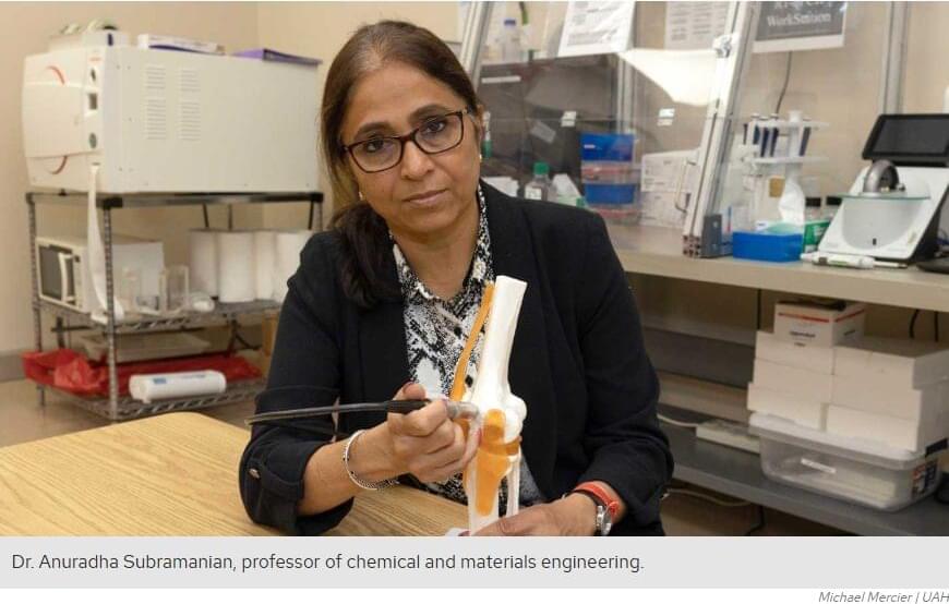
Novel porous gel changes color, shrinks and hardens when it detects target molecules

Researchers at Kyoto University and Tohoku University have developed a new porous polymer gel that selectively recognizes specific molecules (referred to as “guests” in the study) through coordination chemistry and converts these invisible molecular-scale interactions into strikingly visible, macroscale deformation.

The study demonstrates how subtle differences in molecular structure can directly alter the shape, color, and mechanical properties of a soft material, opening new possibilities for “smart” stimuli-responsive materials and molecularly programmable soft matter that can sense and react to its environment.

Molecular recognition is a central concept in supramolecular chemistry and biology, where molecules selectively interact through precisely arranged chemical interactions. While most artificial molecular recognition systems rely on noncovalent interactions such as hydrogen bonding, the present study instead exploits coordination interactions —a type of chemical “handshake”—between metal centers and electron-rich guest molecules.

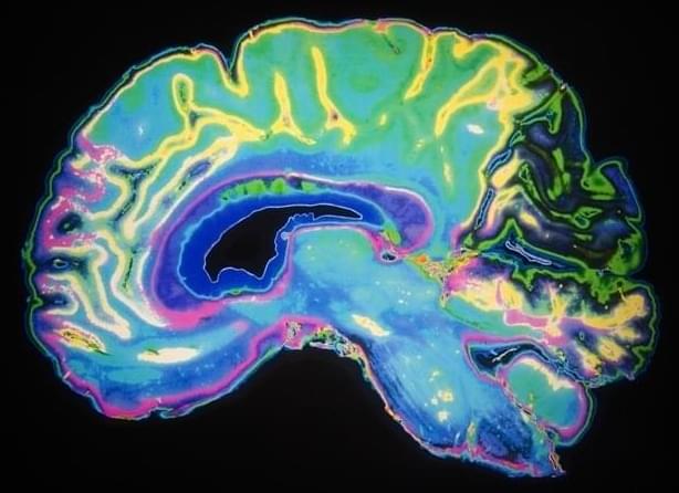
How schizophrenia risk may begin: Gene changes reshape signaling in developing neurons

Researchers at King’s College London have identified the biological nature and timing of changes in human cortical neurons caused by altering activity of a schizophrenia-associated gene in developing human neurons. This discovery links a genetic risk factor to cellular changes in neurons; an essential step for understanding the neurobiology of this mental illness and developing future treatments.

Schizophrenia is estimated to be one of the most heritable psychiatric conditions, with a strong developmental aspect. Large-scale human genomic studies have identified many genetic variants which are thought to increase the likelihood of schizophrenia.

However, the link between these genetic risk variants and the underlying neurobiology of schizophrenia is less well understood. Addressing this knowledge gap provides vital information that could ultimately help develop therapies for the disorder.

Optoelectronic synapse shows exceptional photoresponse for neuromorphic vision

Like so much else in nature, the human visual system has both a complex structure and functional efficiency that is difficult for scientists to replicate. The system is both a sensor and a processor, with the eyes and the brain working together to resolve images with less energy use than anything people have invented.

But a technology called optoelectronic synapses can reproduce at least some of the phenomena that make human vision so successful, and a team of researchers at the National Laboratory of the Rockies (NLR) has discovered why certain materials perform so well at artificial vision and memory.

In their article “Interlayer Exciton Polarons in Mesoscopic V2O5 for Broadband Optoelectronic Synapses” published in Advanced Functional Materials, the NLR-led research team discovered the source of persistent photoconductivity—a mechanism that mirrors some of the functionality of biological synapses in the eye—for a particular vanadium-oxide material.

What if the direction of a magnet could shape the building blocks of life?

In a new discovery, researchers from the Hebrew University of Jerusalem and the Weizmann Institute of Science have found that something in the direction of a magnetic field can influence how molecules of life behave at the most fundamental level and how early chemical processes linked to life may have unfolded.

The study, published in Chem and led by Prof. Yossi Paltiel (Hebrew University) and Prof. Michal Sharon (Weizmann Institute), shows that tiny differences between atoms (different isotopes) can lead to measurable changes in molecular behavior when combined with an invisible quantum property known as electron spin. Separation of the different isotopes can be achieved by magnetic surfaces.

At the center of the story is L-methionine, an amino acid, a basic building block of life. Like other biological molecules, methionine has a specific “handedness,” meaning it exists in a form that is not identical to its mirror image. This property, called chirality, is a mystery: why did nature choose one “hand” over the other?

The quantum key to seeing through chaos

Researchers from the Institut des NanoSciences de Paris, the Kastler Brossel Laboratory and the University of Glasgow have developed an innovative method that renders a scattering medium transparent solely for information carried by entangled photon pairs, while the same medium remains completely opaque to classical light.

Their works are published in the journals Optica (optimization) and Nature Physics (selective image transmission).

Faithfully transmitting spatial information, such as the image of an object, is a major challenge in modern optics. However, this task becomes complex as soon as light travels through disordered media, such as biological tissues, atmospheric turbulence, or multimode optical fibers. In these environments, scattering scrambles the information, making the final image completely unreadable.

/* */