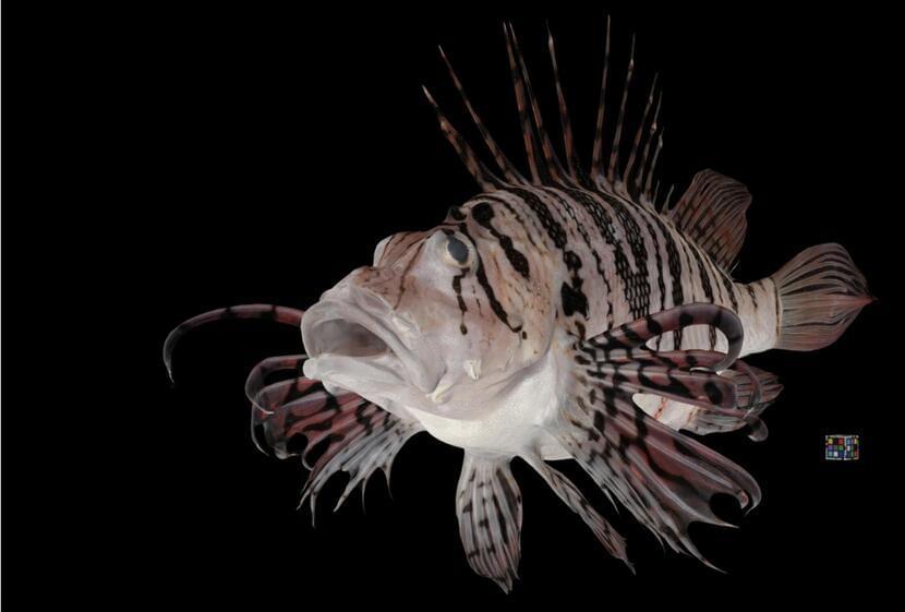
Reporting in Research Ideas and Outcomes, a Kyushu University researcher has developed a new technique for scanning various plants and animals and reconstructing them into highly detailed 3D models. To date, over 1,400 models have been made available online for public use.
Open any textbook or nature magazine and you will find stunning high-resolution pictures of the diverse flora and fauna that encompass our world. From the botanical illustrations in Dioscorides’ De materia medica (50−70 CE) to Robert Hooke’s sketches of the microscopic world in Micrographia (1665), scientists and artists alike have worked meticulously to draw the true majesty of nature.
The advent of photography has given us even more detailed images of animals and plants both big and small, in some cases providing new information on an organism’s morphology. As technology developed, digital libraries began to grow, giving us near unfettered access to valuable data, with methods like computer tomography, or CT, and MRI scanning becoming powerful tools for studying the internal structure of such creatures.


Comments are closed.