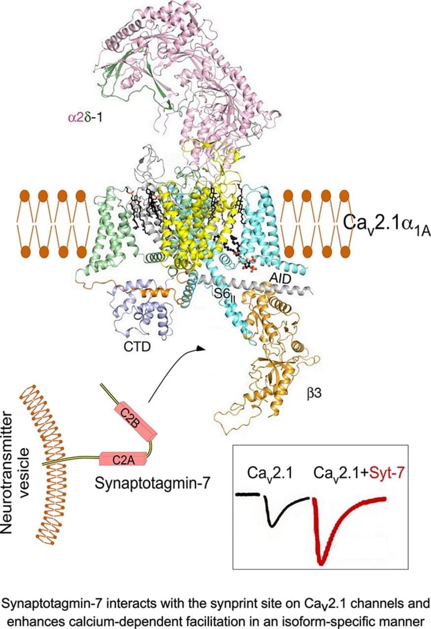
Inward Ca2+ currents conducted by voltage-gated Ca2+ (Cav) channels couple action potentials and other depolarizing stimuli to many Ca2+-dependent intracellular processes, including neurotransmission, hormone secretion, and muscle contraction (Zamponi et al., 2015). In presynaptic nerve terminals, Cav2.1, Cav2.2, and Cav2.3 channels conduct P/Q-type, N-type, and R-type Ca2+ currents that trigger rapid neurotransmission (for review, see Olivera et al., 1994; Zamponi et al., 2015; Nanou and Catterall, 2018). However, only P/Q-type Ca2+ currents conducted by Cav2.1 channels can mediate short-term synaptic facilitation at the calyx of Held in mice (Inchauspe et al., 2004), pointing to a unique role of these Ca2+ channels in short-term synaptic plasticity.
In transfected nonneuronal cells, Ca2+ entry mediated by Cav2.1 channels causes calcium-dependent facilitation (CDF) and inactivation (CDI) during single depolarizations and in trains of repetitive depolarizing pulses (Lee et al., 1999, 2000; DeMaria et al., 2001; Catterall and Few, 2008; Christel and Lee, 2012; Ben-Johny and Yue, 2014). Both CDF and CDI of Cav2.1 channels are dependent on calmodulin (CaM; Lee et al., 1999, 2000; DeMaria et al., 2001). CaM preassociates with the C-terminal domain of the pore-forming α1 subunit of Cav2.1 channels (Erickson et al., 2001). Following Ca2+ binding, CaM initially interacts with the nearby IQ-like motif (IM) and causes CDF, whereas further binding of Ca2+/CaM to the more distal CaM-binding domain (CBD) induces CDI of Cav2.1 channels (DeMaria et al., 2001; Lee et al., 2003). Introducing the IM-AA mutation into the IQ-like motif of Cav2.


Comments are closed.