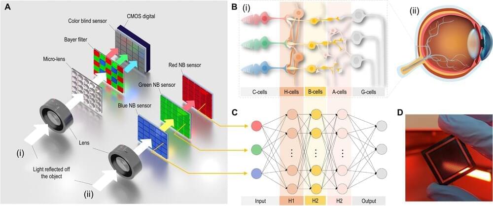
The mammalian retina is a complex system consisting out of cones (for color) and rods (for peripheral monochrome) that provide the raw image data which is then processed into successive layers of neurons before this preprocessed data is sent via the optical nerve to the brain’s visual cortex. In order to emulate this system as closely as possible, researchers at Penn State University have created a system that uses perovskite (methylammonium lead bromide, MAPbX3) RGB photodetectors and a neuromorphic processing algorithm that performs similar processing as the biological retina.
Panchromatic imaging is defined as being ‘sensitive to light of all colors in the visible spectrum’, which in imaging means enhancing the monochromatic (e.g. RGB) channels using panchromatic (intensity, not frequency) data. For the retina this means that the incoming light is not merely used to determine the separate colors, but also the intensity, which is what underlies the wide dynamic range of the Mark I eyeball. In this experiment, layers of these MAPbX3 (X being Cl, Br, I or combination thereof) perovskites formed stacked RGB sensors.
The output of these sensor layers was then processed in a pretrained convolutional neural network, to generate the final, panchromatic image which could then be used for a wide range of purposes. Some applications noted by the researchers include new types of digital cameras, as well as artificial retinas, limited mostly by how well the perovskite layers scale in resolution, and their longevity, which is a long-standing issue with perovskites. Another possibility raised is that of powering at least part of the system using the energy collected by the perovskite layers, akin to proposed perovskite-based solar panels.


