Mar 28, 2023
How to Grow Cerebral Organoids from Human Pluripotent Stem Cells
Posted by Dan Breeden in categories: biotech/medical, education, neuroscience
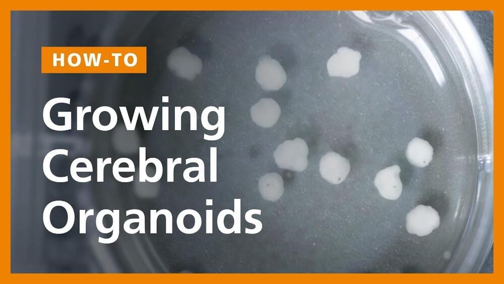
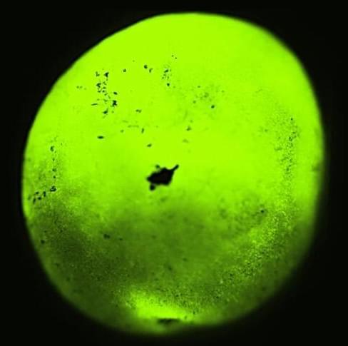
Follow our step-by-step video guide for growing cerebral organoids, or brain organoids, from human pluripotent stem cells (hPSCs). We’ll walk you through embryoid body formation, induction, expansion, and organoid maturation.
0:35 — Embryoid Body Formation.
2:57 — Induction.
4:07 — Expansion.
6:42 — Organoid Maturation.
Continue reading “How to Grow Cerebral Organoids from Human Pluripotent Stem Cells” »