Imagine going for an MRI scan of your knee. This scan measures the density of water molecules present in your knee, at a resolution of about one cubic millimeter – which is great for determining whether, for example, a meniscus in the knee is torn. But what if you need to investigate the structural data of a single molecule that’s five cubic nanometers, or about ten trillion times smaller than the best resolution current MRI scanners are capable of producing? That’s the goal for Dr. Amit Finkler of the Weizmann Institute of Science’s Chemical and Biological Physics Department.
In a recent study (Physical Review Applied, “Mapping Single Electron Spins with Magnetic Tomography”), Finkler, PhD student Dan Yudilevich and their collaborators from the University of Stuttgart, Germany, have managed to take a giant step in that direction, demonstrating a novel method for imaging individual electrons. The method, now in its initial stages, might one day be applicable to imaging various kinds of molecules, which could revolutionize the development of pharmaceuticals and the characterization of quantum materials.
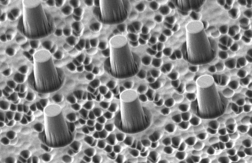
The experimental set-up: A 30-micron-thick diamond membrane with one sensor, on average, at the top of each column, magnified 2,640 times (top) and 32,650 times (bottom)


