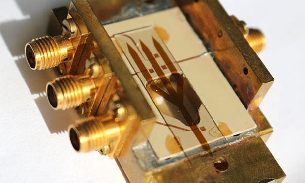
New type of logic gate promises to completely replace electricity with magnetic spin waves for computation.
Faced with the rising threat of drug resistant pathogens, researchers are all but giving up hope that new treatments can be easily cooked up in the lab.
One recent discovery gives us hope that novel antibiotics are easier to find than we think, perhaps in plain sight right under our very feet. And it’s possible their curative properties could have already been recognised centuries in the past.
An international team of researchers based at Swansea University Medical School in South West Wales recently identified a new strain of bacterium in ‘healing soil’ taken from a site associated with ancient druidic rituals in Northern Ireland.
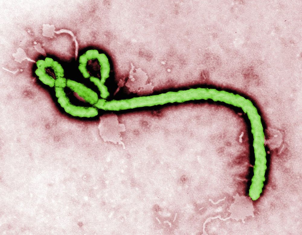
The African country of South Sudan has started vaccinating front-line response staff with the Ebola Zaire vaccine candidate v920.
The vaccine’s producer, Merck, has given 2,160 doses of the vaccine candidate V920 (rVSV-ZEBOV) as part of preparedness measures to fight the spread of the Ebola disease, said the World Health Organization (WHO) in a press release on January 28, 2019.
This preventive effort is in reaction to South Sudan’s neighboring country the Democratic Republic of the Congo (DRC), which is now experiencing its 10th Ebola outbreak.
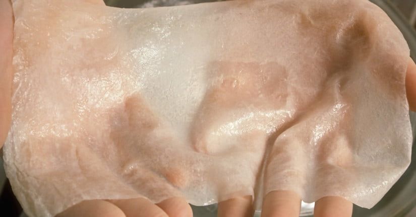
Researchers have developed a new kind of sensor designed to let artificial skin sense pressure, vibrations, and even magnetic fields. Developed by engineers, chemists, and biologists at the University of Connecticut and University of Toronto, the technology could help burn victims and amputees “feel” again through their prosthetic skin.
“The type of artificial skin we developed can be called an electronic skin or e-skin,” Islam Mosa, a postdoctoral fellow at UConn, told Digital Trends. “It is a new group of smart wearable electronics that are flexible, stretchable, shapable, and possess unique sensing capabilities that mimic human skin.”
To create the sensor for the artificial skin, Mosa and his team wrapped a silicone tube with a copper wire and filled the tube with an iron oxide nanoparticle fluid. As the nanoparticles move around the tube, they create an electrical current, which is picked up by the copper wire. When the tube experiences pressure, the current changes.
A patient in Pennsylvania is being tested for a possible Ebola virus infection, according to news reports.
On Wednesday (Feb. 6), the Hospital of the University of Pennsylvania (HUP) said that it was testing the patient for the deadly virus out of “an abundance of caution,” according to local news outlet NBC 10 Philadelphia. Early test results suggest the patient has another condition that is the cause of their illness, but the hospital is taking precautions until the definitive results are in.
“A patient who met screening criteria for Ebola testing is currently being evaluated at HUP while tests to assess the patient’s condition are completed,” Dr. Patrick J. Brennan, chief medical officer at Penn Medicine, told told NBC 10.
Due to their large size, charged surfaces, and environmental sensitivity, proteins do not naturally cross cell-membranes in intact form and, therefore, are difficult to deliver for both diagnostic and therapeutic purposes. Based upon the observation that clustered oligonucleotides can naturally engage scavenger receptors that facilitate cellular transfection, nucleic acid–metal organic framework nanoparticle (MOF NP) conjugates have been designed and synthesized from NU-1000 and PCN-222/MOF-545, respectively, and phosphate-terminated oligonucleotides. They have been characterized structurally and with respect to their ability to enter mammalian cells. The MOFs act as protein hosts, and their densely functionalized, oligonucleotide-rich surfaces make them colloidally stable and ensure facile cellular entry. With insulin as a model protein, high loading and a 10-fold enhancement of cellular uptake (as compared to that of the native protein) were achieved. Importantly, this approach can be generalized to facilitate the delivery of a variety of proteins as biological probes or potential therapeutics.
Proteins play key roles in living systems, and the ability to deliver active proteins to cells is attractive for both diagnostic and therapeutic purposes. Potential uses involve the evaluation of metabolic pathways, regulation of cellular processes, and treatment of disease involving protein deficiencies. (4−6) During the past decade, a series of techniques have been developed to facilitate protein internalization by live cells, including the use of complementary transfection agents, nanocarriers, (7−9) and protein surface modifications. (10−13) Although each strategy has its own merit, none are perfect solutions; they can cause cytotoxicity, reduce protein activity, and suffer from low delivery payloads. (14) For example, we have made the observation that one can take almost any protein and functionalize its surface with DNA to create entities that will naturally engage the cell-surface receptors involved in spherical nucleic acid (SNA) uptake. (13,15−17) While this method is extremely useful in certain situations, it requires direct modification of the protein and large amounts of nucleic acid, on a per-protein basis, to effect transfection. Ideally, one would like to deliver intact, functional proteins without the need to chemically modify them, and to do so in a nucleic-acid efficient manner.
Metal organic frameworks (MOFs) have emerged as a class of promising materials for the immobilization and storage of functional proteins. (18) Their mesoporous structures allow for exceptionally high protein loadings, and their framework architectures can significantly improve the thermal and chemical stabilities of the encapsulated proteins. (19−24) However, although MOF NPs have been recognized as potentially important intracellular delivery vehicles for proteins, (25−27) their poor colloidal stability and positively charged surfaces, (28,29) inhibit their cellular uptake and have led to unfavorable bioavailabilities. (30−33) Therefore, the development of general approaches for reducing MOF NP aggregation, minimizing positive charge (which can cause cytotoxicity), and facilitating cellular uptake is desirable. (34,35)
The “CSI Effect” has been described as being an increased expectation from jurors that forensic evidence will be presented in court that is instantaneous and unequivocal because that is how it is often presented for dramatic effect in television programs and movies. Of course, in reality forensic science, while exact in some respects is just as susceptible to the vagaries of measurements and analyses as any other part of science. In reality, crime scene investigators often spend seemingly inordinate amounts of time gathering and assessing evidence and then present it as probabilities rather than the kind of definitive result expected of a court room filled with actors rather than real people.
Forensics is on the cusp of a third revolution in its relatively young lifetime. The first revolution, under the brilliant but complicated mind of J. Edgar Hoover, brought science to the field and was largely responsible for the rise of criminal justice as we know it today. The second, half a century later, saw the introduction of computers and related technologies in mainstream forensics and created the subfield of digital forensics.
We are now hurtling headlong into the third revolution with the introduction of Artificial Intelligence (AI) – intelligence exhibited by machines that are trained to learn and solve problems. This is not just an extension of prior technologies. AI holds the potential to dramatically change the field in a variety of ways, from reducing bias in investigations to challenging what evidence is considered admissible.
AI is no longer science fiction. A 2016 survey conducted by the National Business Research Institute (NBRI) found that 38% of enterprises are already using AI technologies and 62% will use AI technologies by 2018. “The availability of large volumes of data—plus new algorithms and more computing power—are behind the recent success of deep learning, finally pulling AI out of its long winter,” writes Gil Press, contributor to Forbes.com.
A prototype quantum radar that has the potential to detect objects which are invisible to conventional systems has been developed by an international research team led by a quantum information scientist at the University of York.
The new breed of radar is a hybrid system that uses quantum correlation between microwave and optical beams to detect objects of low reflectivity such as cancer cells or aircraft with a stealth capability. Because the quantum radar operates at much lower energies than conventional systems, it has the long-term potential for a range of applications in biomedicine including non-invasive NMR scans.
The research team led by Dr Stefano Pirandola, of the University’s Department of Computer Science and the York Centre for Quantum Technologies, found that a special converter — a double-cavity device that couples the microwave beam to an optical beam using a nano-mechanical oscillator — was the key to the new system.


