Toggle light / dark theme

Get the latest international news and world events from around the world.

Log in for authorized contributors

Gravitational waves leave imprints on light emitted by atoms, theoretical study predicts

Gravitational waves are ripples in spacetime produced by violent cosmic events, such as the merging of black holes. So far, direct detections have relied on measuring tiny distance changes over kilometer-scale instruments. In a new theoretical study published in Physical Review Letters, researchers at Stockholm University, Nordita, and the University of Tübingen propose an unconventional approach: tracking how gravitational waves reshape the light emitted by atoms. The work describes a possible detection route, but an experimental demonstration remains for the future.

When atoms are excited, they naturally relax by emitting light at a characteristic frequency—a quantum process known as spontaneous emission. This happens through their interaction with the quantum electromagnetic field.

Gravitational waves modulate the quantum field, which in turn affects spontaneous emission,” said Jerzy Paczos, a Ph.D. student at Stockholm University. “This modulation can shift the frequencies of emitted photons compared with the no-wave case.”

Synaptic connectivity alone can reveal neuron types

Recent technological advances facilitate the reconstruction of complete brain connectomes in small organisms and partial connectomes in mammals, involving the mapping of the network of neurons and synaptic connections. Accurate cell typing of these connectomes aids in interpreting circuit functions and comparing brain organization across species.

Traditionally, cell typing relied on manual morphological classification by experts—a slow process that required detailed anatomical information. However, morphology can be deceptive or inadequate in many brain regions, especially in circuits with repeated cell types, where neurons can share very similar morphology despite differing in connectivity.

New X-ray vision for electronics lets scientists monitor working chips remotely

A team of international researchers have developed a breakthrough way to observe what is happening inside electronic chips while they are operating—without touching them, taking them apart, or switching them off. The new technique uses terahertz waves, a safe and non-ionizing form of electromagnetic radiation, to detect tiny movements of electrical charge inside fully packaged semiconductor devices. For the first time, this allows scientists and engineers to monitor electronic components as they function in the real world.

The study, published in the IEEE Journal of Microwaves, involves researchers from Adelaide University in Australia, US technology company Virginia Diodes Inc, the Hasso Plattner Institute and the University of Potsdam, Germany.

Adelaide University Group Leader of the Terahertz Engineering Laboratory (TEL), Professor Withawat Withayachumnankul, said that semiconductors underpin almost every modern technology, from smartphones and medical devices to vehicles, power grids and defense systems.

Frontiers: Information storage and transfer in the brain require a high computational power

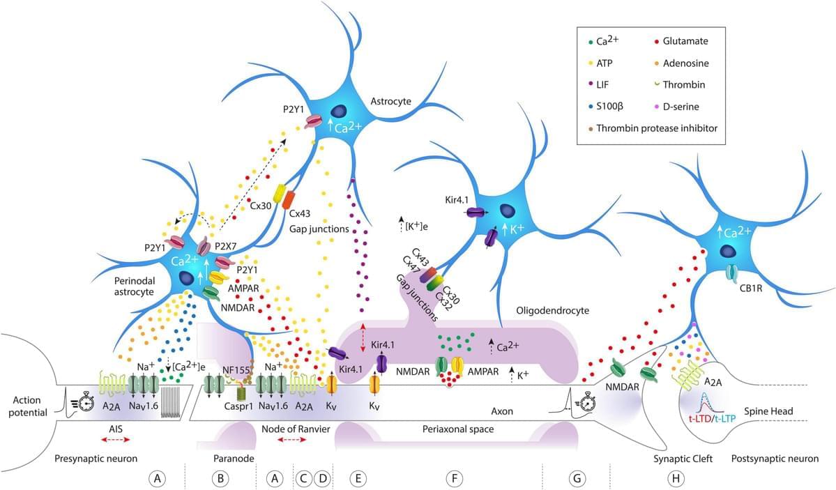
Neuronal network display various local or global mechanisms to allow information storage and transfer in the brain. From synaptic to intrinsic plasticity, the rules of input–output function modulation have been well characterized in neurons. In the past years, astrocytes have been suggested to increase the computational power of the brain and we are only just starting to uncover their role in information processing. Astrocytes maintain a close bidirectional communication with neurons to modify neuronal network excitability, transmission, axonal conduction, and plasticity through various mechanisms including the release of gliotransmitters or local ion homeostasis. Astrocytes have been significantly studied in the context of long-term or short-term synaptic plasticity, but this is not the only mechanism involved in memory formation. Plasticity of intrinsic neuronal excitability also participates in memory storage through regulation of voltage-gated ion channels or axonal morphological changes. Yet, the contribution of astrocytes to these other forms of non-synaptic plasticity remains to be investigated. In this review, we summarized the recent advances on the role of astrocytes in different forms of plasticity and discuss new directions and ideas to be explored regarding astrocytes-neuronal communication and regulation of plasticity.

The rules governing changes in synaptic and intrinsic plasticity are diverse and complex, sometimes synergistic and sometimes not (Debanne et al., 2019). Most studies have been neuro-centric, despite growing evidence that astrocytes can intervene or interact to modify or modulate synaptic transmission (Araque et al., 1998; Jourdain et al., 2007; Bonansco et al., 2011), input integration, neuronal excitability (Tan et al., 2017), spike waveform or axonal conductivity (Sasaki et al., 2011; Lezmy et al., 2021). Astrocytes can detect neuronal activity, and depending on the firing rate of action potentials (APs), they can not only release gliotransmitters such as adenosine or glutamate (Hamilton et al., 2008; Lezmy et al., 2021), but also trigger intracellular calcium ([Ca2+]i) oscillations at different frequencies (Pasti et al., 1997).

From pathology image to biological discovery: LazySlide uses foundation models to connect tissue images and RNA data

Microscopic images of human tissue are a cornerstone of biomedical research and clinical diagnostics. Yet despite their importance, these images often remain difficult to analyze systematically and to connect with other types of biological data. A new study led by CeMM Principal Investigator André Rendeiro and published in Nature Methods introduces “LazySlide,” an open-source software tool that brings the power of foundation models and aims to democratize digital pathology analysis.

Whether it’s an inflamed artery, a tumor spreading into the lung or subtle damage in an organ, when doctors or researchers want to understand what’s happening inside a tissue, one of the most trusted tools is still the microscope. Today, they have largely gone digital: A single tissue sample can be scanned into a whole-slide image so detailed that one can zoom from a bird’s-eye view of the entire tissue down to individual cells. These images, therefore, contain enormous information about tissues from different scales.

However, these images are huge, complex, and often difficult to analyze in a modern, data-driven way. While genetics and single-cell biology have developed effective ways for sharing and comparing data, digital pathology images are hard to incorporate—stored in proprietary formats, processed with incompatible tools, and hard to connect to molecular information like RNA sequencing. Thus, the valuable resources of digitalized tissue images are largely underutilized in many research and clinical settings.

Ultra-thin MoSe₂ grating traps infrared light in a 40-nanometer layer

Controlling light at the micro- and nanoscale opens up opportunities for a better understanding of the world and the development of technology. As modern electronics approaches the limits of its capabilities, photonics comes into play. Instead of manipulating relatively heavy and slow electrons, we can use light and fast photons to encode information. This will make it possible to create devices that are not only faster but also even smaller than those currently in use.

Protein modification in neurodegenerative diseases

The graphical abstract showcases the diversity of posttranslational modifications (PTMs) influencing protein structure and function. It features schematic representations of the following 10 prominent PTMs: phosphorylation (addition of phosphate groups), acetylation (addition of acetyl groups), methylation (addition of methyl groups), SUMOylation (attachment of SUMO proteins), ubiquitylation (attachment of ubiquitin molecules), succinylation (addition of succinyl groups), S-nitrosylation (attachment of NO), ADP-ribosylation (addition of ADP-ribose groups), glycosylation (addition of sugar molecules), and palmitoylation (attachment of palmitate groups).

AI chatbots’ tendency to always agree may reinforce delusions in vulnerable users

The integration of large language model-based AI chatbots into multiple facets of our everyday lives has opened us up to advantages that would have been considered impossible even a decade ago. The same development has, however, opened us up to unforeseen risks, including the impact that engaging with AI chatbots can have on people dealing with mental illness.

AI chatbots are designed to keep conversations going, often by agreeing with users. A article by researchers from King’s College, London, found that this sycophantic tendency may sometimes do more harm than good, reinforcing unusual thoughts rather than challenging them, and potentially contributing to AI-associated delusions, in which users develop or worsen false beliefs about reality.

These interactions can reinforce or even shape delusional beliefs, such as thinking one is uniquely important, being targeted by others, or being in a romantic relationship that does not exist.

Locus coeruleus–amygdala circuit disrupts prefrontal control to impair fear extinction

One of the most-viewed PNAS articles in the last week is “Locus coeruleus–amygdala circuit disrupts prefrontal control to impair fear extinction.” Explore the article here: https://ow.ly/yFH250Ywubb.

For more trending articles, visit https://ow.ly/tZsG50Ywubg.


Stress undermines extinction learning and hinders exposure-based clinical therapies for a variety of neuropsychiatric disorders. In both animals and humans, dysfunction in the ventromedial prefrontal cortex (vmPFC) contributes to stress-impaired extinction, but the neural circuit by which stress modulates vmPFC function is not known. We hypothesize that locus coeruleus (LC) norepinephrine undermines extinction learning by recruiting projections from the basolateral amygdala (BLA) to vmPFC. Using a combination of circuit-specific chemogenetics and calcium imaging, we find that activation of LC noradrenergic neurons mimics a behavioral stressor (footshock), induces freezing behavior, reduces spontaneous neuronal activity in the vmPFC, impairs extinction learning, and alters the population dynamics of vmPFC ensembles.

/* */