Toggle light / dark theme

Get the latest international news and world events from around the world.

Log in for authorized contributors

How deadly Marburg virus enters human cells

The researchers also discovered a tiny antibody, called a nanobody, which mimics NPC1 at the receptor-binding site and can slip past a protective cap on Marburg’s entry protein, bind to it and block its attachment to the receptor. In lab tests, this nanobody prevented Marburg virus from entering cells. ScienceMission sciencenewshighlights.


In a new study published in Nature the researchers found that the Marburg virus (MBV), one of the world’s deadliest pathogens with an average 73% fatality rate, is unusually efficient at getting inside human cells. They also showed that the virus’s entry protein contains structural features that explain this efficiency and point to a strategy for blocking infection.

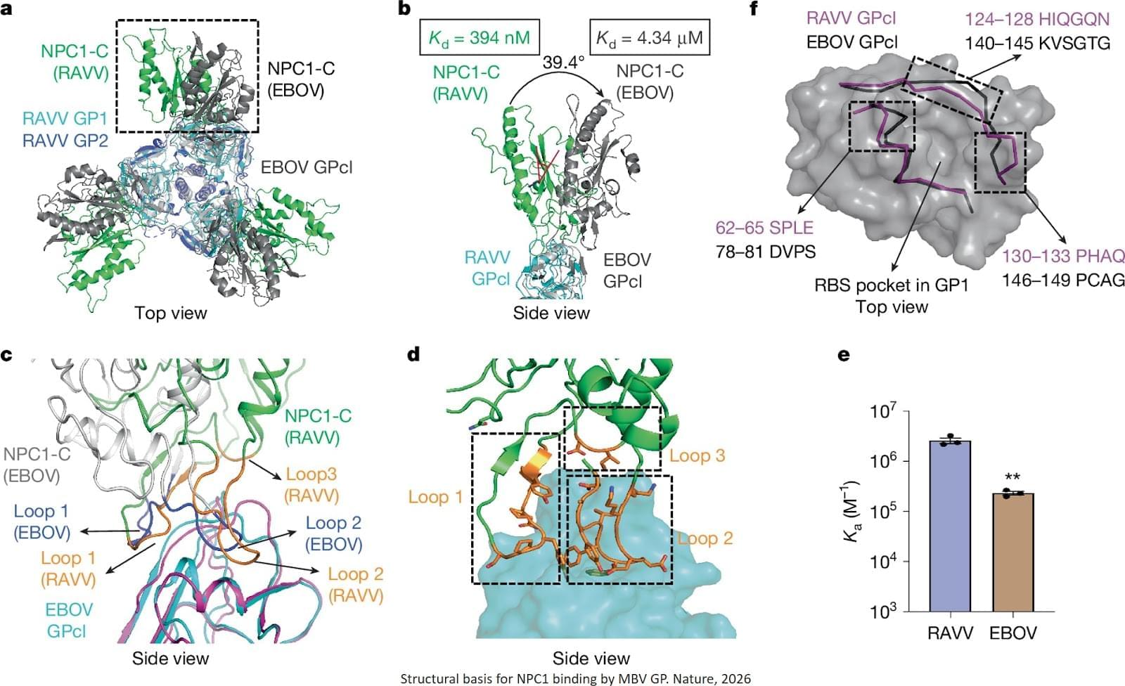
The researchers designed a tightly controlled system that enables a fair comparison of the entry proteins of Marburg and its relative Ebola. The team further found that the two viruses share the same human receptor. The authors determined structures of MBV glycoprotein (GP) in three states: unbound; bound to its endosomal receptor NPC1; and complexed with a neutralizing nanobody.

Marburg’s entry protein binds this receptor in a distinct orientation and with higher affinity, then changes shape in ways that help the virus enter cells. The authors show that the glycan cap shields the receptor-binding site from NPC1 but only partially from the nanobody, enabling limited immune evasion. After glycan cap cleavage, NPC1 binds to MBV GP in a distinct orientation compared with EBOV GP, providing an additional anchor and enhancing receptor affinity. NPC1 engagement also induces substantial conformational changes in MBV GP, probably facilitating membrane fusion. Using this approach, they showed that Marburg’s entry protein can drive viral entry into human cells up to 300 times more efficiently than Ebola’s.

10 Ancient Space Objects That Existed Before the Universe Itself

All right, let’s go.
Number 10. Methuselah’s Star.
In 2000, a team of astronomers led by Howard Bond at Penn State University pointed the Hubble Space Telescope at a faint star in the constellation Libra and made a discovery that should have been impossible. The star, designated HD 140,283 and later nicknamed Methuselah, appeared to be 14.5 billion years old. The universe itself is only 13.8 billion years old. A star older than the cosmos that contains it shouldn’t exist. Yet there it was, burning quietly just 190 light years from Earth, defying the most fundamental timeline in all of physics.

Gödel Handed Einstein a Universe With Time Travel

Take your personal data back with Incogni! Use code CURT at the link below and get 60% off an annual plan: https://incogni.com/curt.

As a listener of TOE you can get a special 20% off discount to The Economist and all it has to offer! Visit https://www.economist.com/toe.

FOLLOW:
Spotify: https://open.spotify.com/show/4gL14b9

  • Substack: https://curtjaimungal.substack.com/su
  • Twitter: / toewithcurt
  • Discord Invite: / discord
  • Crypto: https://commerce.coinbase.com/checkou
  • PayPal: https://www.paypal.com/donate?hosted_
  • Guests do not pay to appear. #science.
    Substack: https://curtjaimungal.substack.com/su
    Twitter: / toewithcurt.
    Discord Invite: / discord.
    Crypto: https://commerce.coinbase.com/checkou
    PayPal: https://www.paypal.com/donate?hosted_

    Guests do not pay to appear. #science

    Read more

    The Physicist Who Proved Entropy = Gravity

    What if gravity is not fundamental but emerges from quantum entanglement? In this episode, physicist Ted Jacobson reveals how Einstein’s equations can be derived from thermodynamic principles of the quantum vacuum, reshaping our understanding of space, time, and gravity itself.

    As a listener of TOE you can get a special 20% off discount to The Economist and all it has to offer! Visit https://www.economist.com/toe.

    Join My New Substack (Personal Writings): https://curtjaimungal.substack.com.

    Listen on Spotify: https://tinyurl.com/SpotifyTOE

    Become a YouTube Member (Early Access Videos):
    / @theoriesofeverything.

    Timestamps:

    Genius 10 Year Old’s Research Shocks Scientists Around the World

    How do we evolve/survive?


    At just 10 years old, Jo Nagai conducted an experiment that would surprise scientists around the world.

    By raising and training swallowtail caterpillars at home, Jo demonstrated that butterflies can retain memories formed during their larval stage, even after undergoing complete metamorphosis. But what he discovered next was even more unexpected: those learned responses appeared to persist to the next generation.

    This video explores Jo’s experiment, the scientific ideas behind it, and the simple question that started it all.

    #research #science #nature #animals #animalbehavior #discovery #butterfly.

    Gravitational waves leave imprints on light emitted by atoms, theoretical study predicts

    Gravitational waves are ripples in spacetime produced by violent cosmic events, such as the merging of black holes. So far, direct detections have relied on measuring tiny distance changes over kilometer-scale instruments. In a new theoretical study published in Physical Review Letters, researchers at Stockholm University, Nordita, and the University of Tübingen propose an unconventional approach: tracking how gravitational waves reshape the light emitted by atoms. The work describes a possible detection route, but an experimental demonstration remains for the future.

    When atoms are excited, they naturally relax by emitting light at a characteristic frequency—a quantum process known as spontaneous emission. This happens through their interaction with the quantum electromagnetic field.

    Gravitational waves modulate the quantum field, which in turn affects spontaneous emission,” said Jerzy Paczos, a Ph.D. student at Stockholm University. “This modulation can shift the frequencies of emitted photons compared with the no-wave case.”

    Synaptic connectivity alone can reveal neuron types

    Recent technological advances facilitate the reconstruction of complete brain connectomes in small organisms and partial connectomes in mammals, involving the mapping of the network of neurons and synaptic connections. Accurate cell typing of these connectomes aids in interpreting circuit functions and comparing brain organization across species.

    Traditionally, cell typing relied on manual morphological classification by experts—a slow process that required detailed anatomical information. However, morphology can be deceptive or inadequate in many brain regions, especially in circuits with repeated cell types, where neurons can share very similar morphology despite differing in connectivity.

    New X-ray vision for electronics lets scientists monitor working chips remotely

    A team of international researchers have developed a breakthrough way to observe what is happening inside electronic chips while they are operating—without touching them, taking them apart, or switching them off. The new technique uses terahertz waves, a safe and non-ionizing form of electromagnetic radiation, to detect tiny movements of electrical charge inside fully packaged semiconductor devices. For the first time, this allows scientists and engineers to monitor electronic components as they function in the real world.

    The study, published in the IEEE Journal of Microwaves, involves researchers from Adelaide University in Australia, US technology company Virginia Diodes Inc, the Hasso Plattner Institute and the University of Potsdam, Germany.

    Adelaide University Group Leader of the Terahertz Engineering Laboratory (TEL), Professor Withawat Withayachumnankul, said that semiconductors underpin almost every modern technology, from smartphones and medical devices to vehicles, power grids and defense systems.

    Frontiers: Information storage and transfer in the brain require a high computational power

    Neuronal network display various local or global mechanisms to allow information storage and transfer in the brain. From synaptic to intrinsic plasticity, the rules of input–output function modulation have been well characterized in neurons. In the past years, astrocytes have been suggested to increase the computational power of the brain and we are only just starting to uncover their role in information processing. Astrocytes maintain a close bidirectional communication with neurons to modify neuronal network excitability, transmission, axonal conduction, and plasticity through various mechanisms including the release of gliotransmitters or local ion homeostasis. Astrocytes have been significantly studied in the context of long-term or short-term synaptic plasticity, but this is not the only mechanism involved in memory formation. Plasticity of intrinsic neuronal excitability also participates in memory storage through regulation of voltage-gated ion channels or axonal morphological changes. Yet, the contribution of astrocytes to these other forms of non-synaptic plasticity remains to be investigated. In this review, we summarized the recent advances on the role of astrocytes in different forms of plasticity and discuss new directions and ideas to be explored regarding astrocytes-neuronal communication and regulation of plasticity.

    The rules governing changes in synaptic and intrinsic plasticity are diverse and complex, sometimes synergistic and sometimes not (Debanne et al., 2019). Most studies have been neuro-centric, despite growing evidence that astrocytes can intervene or interact to modify or modulate synaptic transmission (Araque et al., 1998; Jourdain et al., 2007; Bonansco et al., 2011), input integration, neuronal excitability (Tan et al., 2017), spike waveform or axonal conductivity (Sasaki et al., 2011; Lezmy et al., 2021). Astrocytes can detect neuronal activity, and depending on the firing rate of action potentials (APs), they can not only release gliotransmitters such as adenosine or glutamate (Hamilton et al., 2008; Lezmy et al., 2021), but also trigger intracellular calcium ([Ca2+]i) oscillations at different frequencies (Pasti et al., 1997).

    From pathology image to biological discovery: LazySlide uses foundation models to connect tissue images and RNA data

    Microscopic images of human tissue are a cornerstone of biomedical research and clinical diagnostics. Yet despite their importance, these images often remain difficult to analyze systematically and to connect with other types of biological data. A new study led by CeMM Principal Investigator André Rendeiro and published in Nature Methods introduces “LazySlide,” an open-source software tool that brings the power of foundation models and aims to democratize digital pathology analysis.

    Whether it’s an inflamed artery, a tumor spreading into the lung or subtle damage in an organ, when doctors or researchers want to understand what’s happening inside a tissue, one of the most trusted tools is still the microscope. Today, they have largely gone digital: A single tissue sample can be scanned into a whole-slide image so detailed that one can zoom from a bird’s-eye view of the entire tissue down to individual cells. These images, therefore, contain enormous information about tissues from different scales.

    However, these images are huge, complex, and often difficult to analyze in a modern, data-driven way. While genetics and single-cell biology have developed effective ways for sharing and comparing data, digital pathology images are hard to incorporate—stored in proprietary formats, processed with incompatible tools, and hard to connect to molecular information like RNA sequencing. Thus, the valuable resources of digitalized tissue images are largely underutilized in many research and clinical settings.

    /* */