Toggle light / dark theme

New insights into the epigenetic processes via which neuroinflammation causes memory loss

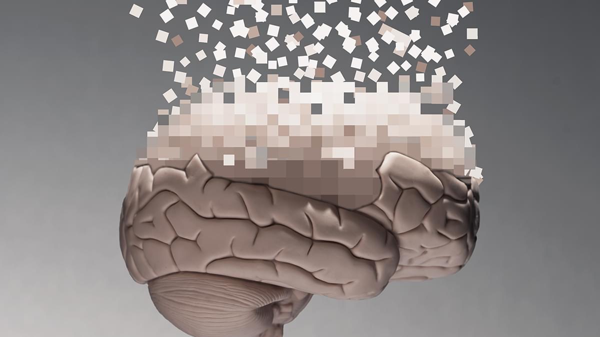
Neuroinflammation, a prolonged activation of the brain’s immune system prompted by infections or other factors, has been linked to the disruption of normal mental functions. Past studies, for instance, have found that neuroinflammation plays a central role in neurodegenerative diseases, medical conditions characterized by the progressive degradation of cells in the spinal cord and brain.

When inflammation is taking place, cells release proteins that act as signals between immune cells, also known as cytokines. While some studies have linked a specific cytokine called interleukin-1 (IL-1) to changes in brain function, the mechanisms through which it could contribute to a decline in mental capabilities remain poorly understood.

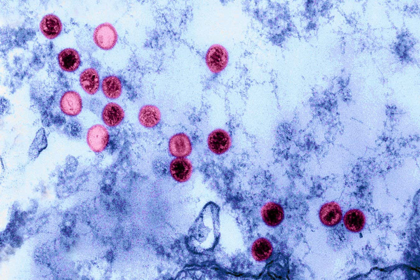
Researchers at the University of Toulouse INSERM and CNRS recently carried out a study involving mice aimed at better understanding these mechanisms. Their paper, published in Nature Neuroscience, particularly focused on neuroinflammation elicited by the parasite Toxoplasma gondii (T. gondii), which is responsible for a well-known illness called toxoplasmosis.

How an autism-linked mutation reduces vasopressin and alters social behavior

A team of researchers has identified for the first time the mechanism linking a mutation in the Shank3 gene with alterations in social behavior. Using a mouse model carrying this autism-associated mutation, the study shows that vasopressin, a brain hormone essential for social relationships, is not properly released in the lateral septum.

The team is from the Cognition and Social Interactions laboratory, led by Félix Leroy at the Institute for Neurosciences, a joint center of the Spanish National Research Council (CSIC) and the Miguel Hernández University (UMH) of Elche.

The work, published in Nature Communications, demonstrates that the proper release of vasopressin in this region regulates social behaviors through two distinct receptor pathways: one controlling sociability and the other controlling social aggression, and that selective activation of these receptors can reverse deficits in social interaction without triggering unwanted aggressive responses.

Apertura Gene Therapy and Rett Syndrome Research Trust Collaborate to Pioneer Advanced Genetic Medicines for Rett Syndrome Using TfR1-Targeted AAV Capsid

NEW YORK and TRUMBULL, Conn., April 30, 2025 /PRNewswire/ — Apertura Gene Therapy, a biotechnology company focused on innovative gene therapy solutions, and the Rett Syndrome Research Trust (RSRT), an organization working to cure Rett Syndrome, today announced a collaboration to license Apertura’s human transferrin receptor 1 capsid (TfR1 CapX). This partnership aims to advance innovative genetic medicine approaches for the treatment of Rett Syndrome, a rare genetic neurological disorder caused by random mutations in the MECP2 gene on the X chromosome that primarily affect females, causing developmental regression and severe motor and language impairments.

Apertura’s TfR1 CapX is an intravenously delivered adeno-associated virus (AAV) capsid engineered to bind the transferrin receptor 1(TfR1), enabling efficient delivery of genetic medicines across the blood-brain barrier (BBB). TfR1 is a well-characterized BBB-crossing receptor, broadly and consistently expressed throughout life—even in the context of neurological disease—making it an attractive target for CNS delivery in disorders like Rett syndrome. Developed by Apertura’s academic founder, Dr. Ben Deverman, Director of Vector Engineering at the Broad Institute, TfR1 CapX has shown strong CNS selectivity in preclinical studies, achieving over 50% neuronal and 90% astrocyte transduction across multiple brain regions. Because Rett syndrome affects the brain diffusely, broader cellular transduction may correlate with greater symptomatic improvement.

SeeMe detects hidden signs of consciousness in brain injury patients

SeeMe, a computer vision tool tested by Stony Brook University researchers, was able to detect low-amplitude, voluntary facial movements in comatose acute brain injury patients days before clinicians could identify overt responses.

Close friends know that I have a standing “do not unplug” order should I ever fall into an unresponsive state. If there is even a flicker of a chance that the mind is still working, I will be fine. Keep me plugged in and hang a “do not disturb” sign on whatever apparatus is keeping me alive.

It’s not like you can know in advance what it’s like, but it seems relaxed enough, with plenty of time to think, and I haven’t really gained anything useful from conversations with other humans in years (aside from my editors who always provide valuable information). If it is at all like sleeping, there might be dreams, so, perchance, that’s what I’d be doing in a comatose state. But for the friends by my bedside, how to be certain that the mind is still flickering?

Brains listen best in the ‘Goldilocks echo zone,’ says study

Macquarie University hearing researchers have discovered how our brains learn to listen, and how this can help us understand speech in noisy, echo-filled spaces.

The research, published in eLife, looks at how we can unconsciously adjust to different kinds of .

Building on earlier work that showed animals’ brains quickly adapt to changes in sound levels, the new study is the first to show how humans adapt to echoey environments to improve their speech understanding.

/* */