The system, which also synthesizes her voice, takes no more than a second to translate thoughts to speech.
Category: neuroscience – Page 77

Mapping brain development at the protein level in unprecedented detail
Researchers at the University of Virginia have created the first comprehensive protein-level atlas of brain development, providing unprecedented insight into how the brain forms and potential implications for understanding neurological disorders. The study, published in Nature Neuroscience, analyzed over 24 million individual cells from mouse brains, revealing detailed molecular pathways that guide brain development from early embryonic stages through early postnatal development.
The research team, led by Professors Christopher Deppmann and Eli Zunder, used an innovative technique called mass cytometry to track 40 different proteins across various brain regions and developmental stages. The approach provided a more detailed view of cellular function than previous studies that primarily examined RNA.
“While RNA studies have given us important insights, proteins are the actual workforce of cells,” explained Deppmann, a professor in the College and Graduate School of Arts & Sciences’ Department of Biology. “By studying proteins directly, we can better understand how cells are functioning and communicating during brain development.”
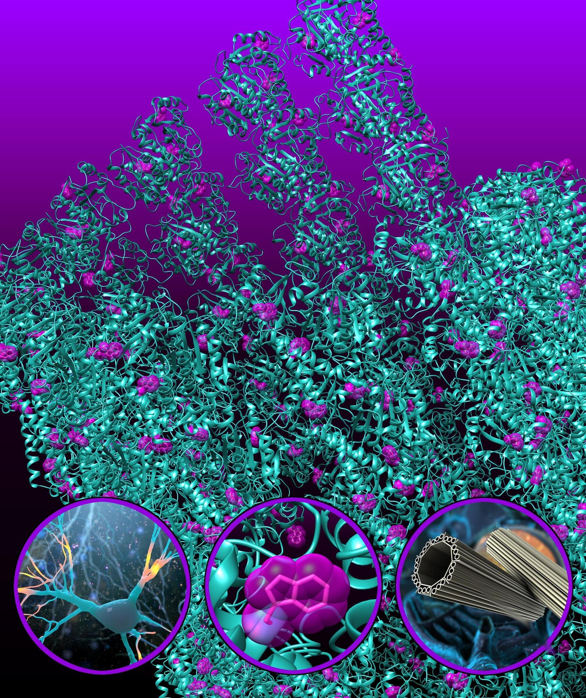
Quantum fiber optics in the brain enhance processing, may protect against degenerative diseases
The effects of quantum mechanics—the laws of physics that apply at exceedingly small scales—are extremely sensitive to disturbances. This is why quantum computers must be held at temperatures colder than outer space, and only very, very small objects, such as atoms and molecules, generally display quantum properties.
By quantum standards, biological systems are quite hostile environments: they’re warm and chaotic, and even their fundamental components—such as cells—are considered very large.
But a group of theoretical and experimental researchers has discovered a distinctly quantum effect in biology that survives these difficult conditions and may also present a way for the brain to protect itself from degenerative diseases like Alzheimer’s.
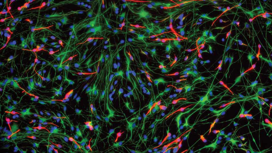
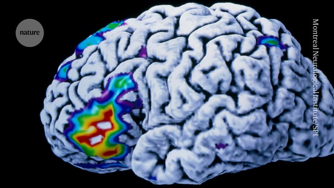

Gold Nanoparticles in Parkinson’s Disease Therapy: A Focus on Plant-Based Green Synthesis
Parkinson’s disease (PD) is a progressive neurodegenerative disease that affects approximately 1% of people over the age of 60 and 5% of those over the age of 85. Current drugs for Parkinson’s disease mainly affect the symptoms and cannot stop its progression. Nanotechnology provides a solution to address some challenges in therapy, such as overcoming the blood-brain barrier (BBB), adverse pharmacokinetics, and the limited bioavailability of therapeutics. The reformulation of drugs into nanoparticles (NPs) can improve their biodistribution, protect them from degradation, reduce the required dose, and ensure target accumulation. Furthermore, appropriately designed nanoparticles enable the combination of diagnosis and therapy with a single nanoagent.
In recent years, gold nanoparticles (AuNPs) have been studied with increasing interest due to their intrinsic nanozyme activity. They can mimic the action of superoxide dismutase, catalase, and peroxidase. The use of 13-nm gold nanoparticles (CNM-Au8®) in bicarbonate solution is being studied as a potential treatment for Parkinson’s disease and other neurological illnesses. CNM-Au8® improves remyelination and motor functions in experimental animals.
Among the many techniques for nanoparticle synthesis, green synthesis is increasingly used due to its simplicity and therapeutic potential. Green synthesis relies on natural and environmentally friendly materials, such as plant extracts, to reduce metal ions and form nanoparticles. Moreover, the presence of bioactive plant compounds on their surface increases the therapeutic potential of these nanoparticles. The present article reviews the possibilities of nanoparticles obtained by green synthesis to combine the therapeutic effects of plant components with gold.

Scientists Just Discovered Quantum Signals Inside Life Itself
Biological systems, once thought too chaotic for quantum effects, may be quietly leveraging quantum mechanics to process information faster than anything man-made.
New research suggests this isn’t just happening in brains, but across all life, including bacteria and plants.
Schrödinger’s legacy inspires a quantum leap.
Short-term reactivation of brain between encoding of memories enhances recall, study finds
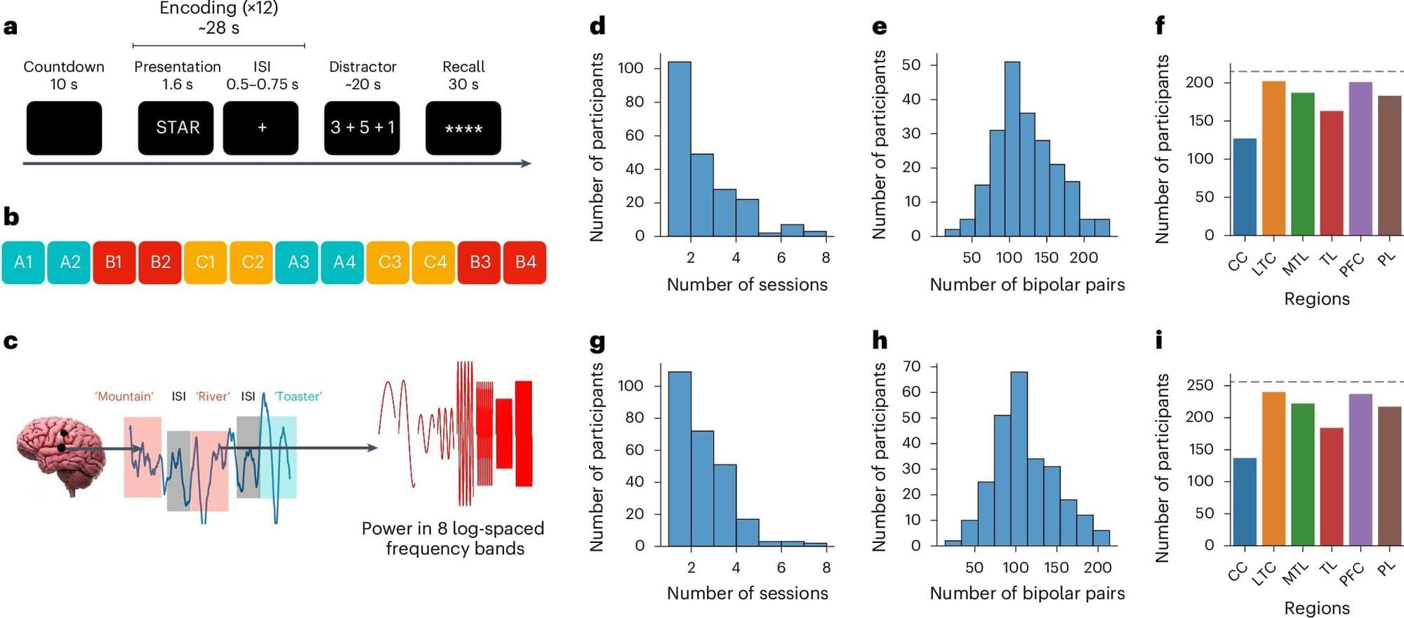
Past neuroscience and psychology studies have shown that after the human brain encodes specific events or information, it can periodically reactivate them to facilitate their retention, via a process known as memory consolidation. The reactivation of memories has been specifically studied in the context of sleep or rest, with findings suggesting that during periods of inactivity, the brain reactivates specific memories, allowing people to remember them in the long term.
Researchers at the University of Pennsylvania and other institutions in the United States recently conducted a study exploring the possibility that the brain engages in a similar reactivation process during wakefulness to store important information for shorter periods of time. Their findings, published in Nature Neuroscience, suggest that the spontaneous reactivation of specific stimuli in the brain during the brief intervals between their encoding predicts the accuracy with which people remember them at the end of a memory task.
“Mike Kahana and I were both quite interested in the long history of thinking about rehearsal and its effects on the way in which people later recalled things,” Dr. David Halpern, the first author of the paper, told Medical Xpress. “Rehearsal is challenging to study since people often do it without any overt behavior (unless we ask them to rehearse out loud).”