Toggle light / dark theme

Interleukin-9 Regulates NF-kB-Mediated Activation of Astrocytes in Multiple Sclerosis Brain

Background and ObjectivesInterleukin-9 (IL-9) is an immune molecule with multiple roles in a variety of cell types. IL-9–induced cell responses are mediated by the IL-9 receptor (IL-9R). Recent evidence demonstrates that expression of IL-9R in post mortem…

A Brain Parasite Infecting Millions Is Far Less Sleepy Than We Thought

A parasite that lives permanently in the brains of millions may not be as uniformly dormant as scientists once thought.

Researchers at the University of California, Riverside (UCR) have recently found evidence of low-level T. gondii reactivation in the brains of mice, even during long-term infection.

Today, more than a third of the world’s human population is infected by Toxoplasma gondii, a brain-invading parasite that reproduces in cats with mice and other animals acting as intermediate hosts.

Neuralink’s Brain Chip: How It Works and What It Means

Elon Musk recently announced that Neuralink, his company aiming to revolutionize brain-computer interfaces (BCIs), has successfully implanted a brain chip in a human for the first time. The implantation of the device, called “the Link,” represents a leap forward in the realm of BCIs, which record and decode brain activity, that may allow for new innovations in health care, communication, and cognitive abilities.

Though limited information on the technology is available and Neuralink’s claims have not been independently verified, here’s a look at the Link, its functionality, and the potential implications of this groundbreaking innovation.

Native postsynaptic density is a functional condensate formed via phase separation

To obtain direct evidence supporting the theory that the postsynaptic density (PSD) in neuronal synapses is formed via phase separation, Chen et al. purified and characterized the native PSD from the mouse brain. Their results demonstrate that the native PSD has characteristic features of biological condensates formed via phase separation.

Procrastination in adulthood linked to brain development during adolescence

Procrastination, the tendency to unnecessarily delay or put off tasks even if this will have negative consequences, is a common behavior for many people. While occasionally delaying or putting off bothersome tasks is not necessarily problematic, severe and prolonged procrastination is closely tied to some neuropsychiatric disorders, including attention-deficit/hyperactivity disorder (ADHD) and anxiety disorders.

Unveiling patterns in the brain’s structure and genetic factors linked to procrastination could help to reliably uncover this tendency to postpone tasks in affected individuals. This could in turn inform the development of preventative strategies or interventions that tackle procrastination early, before it exacerbates other underlying mental health disorders.

Researchers at the Chinese Academy of Sciences and other institutes in China recently carried out a study aimed at shedding new light on the biological and genetic roots of procrastination. Their paper, published in Molecular Psychiatry, outlines specific patterns in the brain’s structure during adolescence that are linked to procrastination in adulthood.

Prenatal exposure to air pollution associated with lower cognitive performance in early childhood

The prenatal period is a critical window for brain development, yet few studies have examined the impact of air pollution exposure during pregnancy on child cognition. A new study led by the Barcelona Institute for Global Health (ISGlobal), in collaboration with the University of Barcelona (UB), shows that prenatal exposure to pollution is associated with lower cognitive performance in newborns.

These findings highlight the importance of reducing air pollution exposure, especially during pregnancy, to protect neurodevelopment.

The study, published in Environmental Pollution, included data from 168 mother-child pairs participating in the BiSC (Barcelona Life Study Cohort) project, conducted in Barcelona between 2018 and 2023.

Brain Scans Reveal an “Inflamed Brain Type” Across Major Psychiatric Disorders

Individuals with psychiatric disorders exhibiting seemingly similar symptoms often respond very differently to the same treatment, suggesting that distinct biological processes are at work beneath the surface of similar clinical presentations. Researchers have now identified a distinct immuno-inflammatory biomarker across major psychiatric disorders that can be detected using non-invasive brain imaging. Patients exhibiting this brain signature showed systemic inflammation and poorer response to standard treatments. The findings of the new study in Biological Psychiatry, published by Elsevier, lay the foundation for a biology-augmented diagnostic framework in psychiatry and detail the potential for biomarker-guided, anti-inflammatory precision therapies.

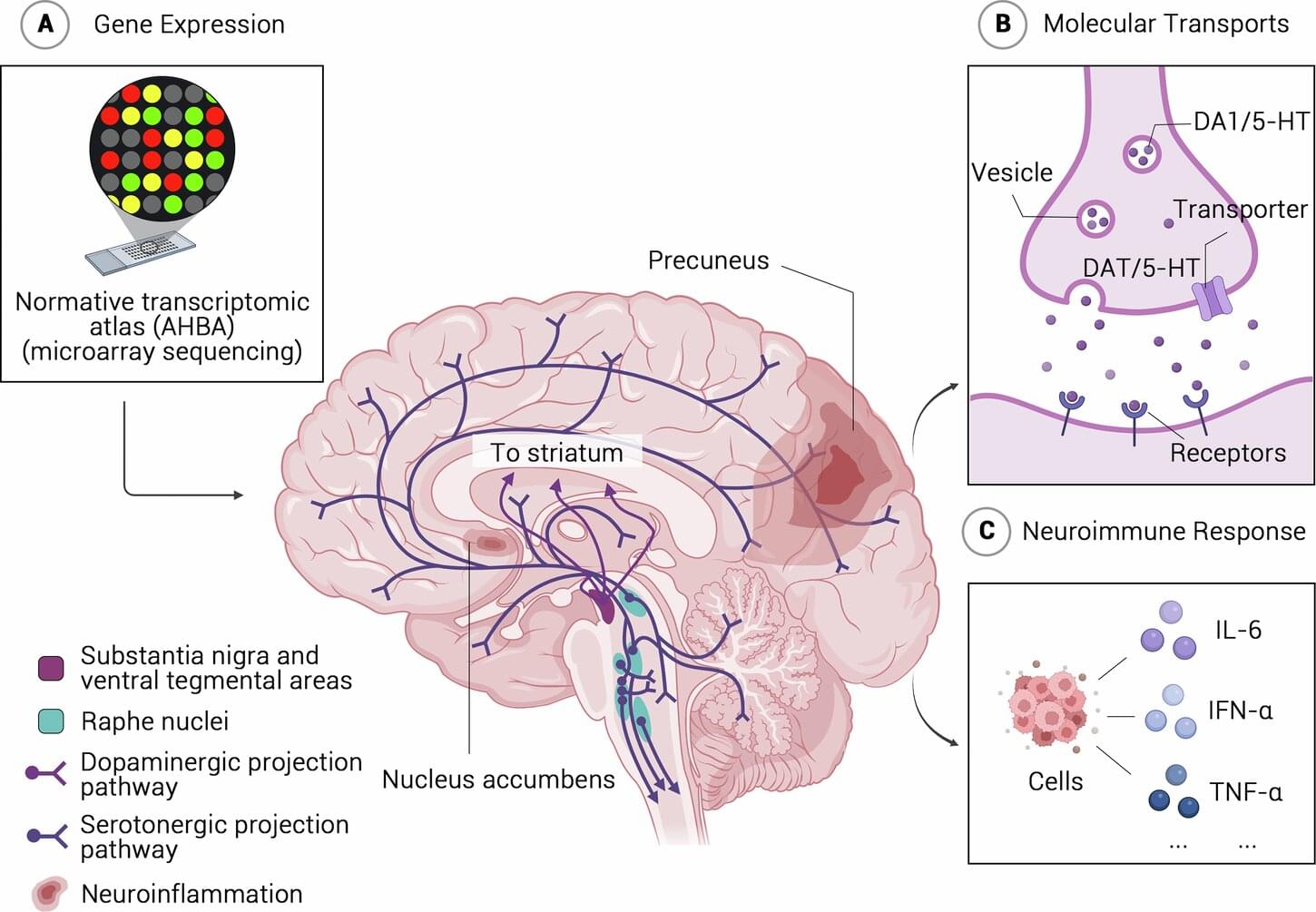
Neuroimaging links diverse biological mechanisms to clinical manifestations, providing compelling insights into the neural mechanisms underlying brain function implicated in psychiatric diseases. Through neuroimaging, shared neural correlates have been increasingly identified across major psychiatric disorders such as schizophrenia, major depressive disorder, and bipolar disorder. While subtypes within and across psychiatric diagnoses have been identified, the biological underpinnings remain unclear. This study aimed to uncover these hidden “biotypes,” focusing particularly on brain inflammation—a mechanism thought to drive illness in a subset of patients, but which is difficult to measure directly in the living brain.

The research was conducted in two independent cohorts. In the first stage, brain connectivity scans were combined with blood-based molecular (DNA methylation) data to identify a brain network pattern linked to immune system dysfunction. In the second longitudinal stage, investigators validated that patients with this brain marker had higher blood inflammation indices—such as neutrophil-to-lymphocyte ratios—and showed less improvement with conventional treatments during hospitalization.

Neuron position found less crucial for brain connectivity than once thought

The human brain contains billions of connected neurons that collectively support different mental functions, including the processing of sensory information, the encoding of memories, attention processes, and decision-making. For a long time, neuroscientists have assumed the position of specific neurons in the brain plays a key role in the brain’s connectivity and proper functioning.

Researchers at University of Geneva, INSERM, Ecole Polytechnique Fédérale de Lausanne and other institutes recently gathered evidence that contradicts this long-standing assumption, showing misplaced neurons can still retain their “identity,” connect with other neurons and support the processing of sensory information.

Their paper, published in Nature Neuroscience, could reshape the present understanding of developmental disorders and other conditions linked to the rearrangement of neurons or cortical malformations.

/* */