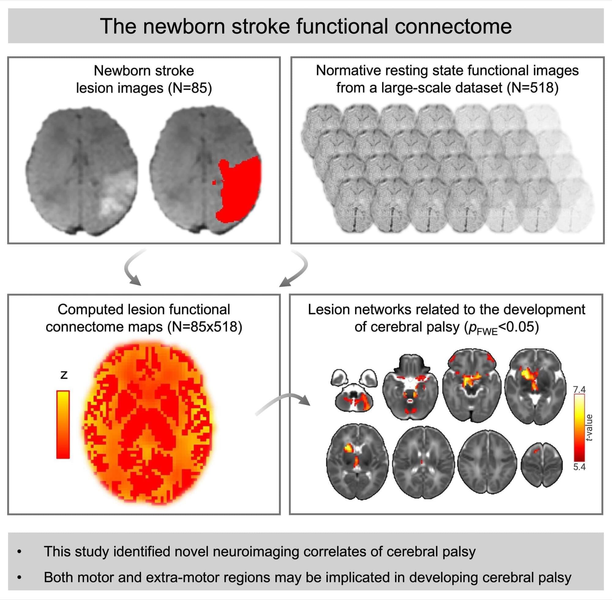
The development of cerebral palsy after neonatal stroke may be associated with disruptions of broad functional networks involving motor and extramotor regions as opposed to isolated lesions of the motor tracts.
Prior studies have established that resting-state networks are already present in newborns as early as term-equivalent age19 and that motor outcomes in healthy populations and other clinical populations are related to both primary motor networks and other broader motor networks.40,41 Our observation that the development of cerebral palsy correlated with both motor and extramotor regions lines up with these findings in other populations, and may indicate more widespread network vulnerabilities after stroke in neonates compared with adults.21 While it is known that disruption to primary motor regions is related to the development of cerebral palsy after NAIS,5,8 the extramotor regions (the frontal and temporal regions identified in the current study) may be more vulnerable to network-level disruptions in neonatal stroke due to the known relative immaturity of these regions during the neonatal period and their prolonged maturation to support higher-order functions.42,43 Particular vulnerability of these temporal and frontal regions has been demonstrated in other newborn, very preterm populations at risk of brain insult and injury.43
There are limitations to this study. Lesion network mapping has frequently been performed in adults and is more clinically feasible than directly acquiring resting-state fMRI scans in patients with NAIS; however, lesion network mapping may be seen as a less direct method for assessing brain connectivity.13,21,22 Functional connectivity has been well characterized in adults and neonates,1,16–19 but future studies will need to examine structural connectivity between regions via white matter fiber pathways using diffusion MRI scans to determine whether the functional changes identified correspond to structural changes.44 Such diffusion MRI investigations will be important in the future to investigate the roles (as part of global structural networks) of white matter regions and tracts such as the posterior limb of the internal capsule, which has previously been shown to be a strong predictor of motor outcome using individual region-based analyses.5–12 Given the paucity of prior research in this area, we aimed to establish that there is a relationship between the lesion functional connectome and cerebral palsy, but future work will also need to investigate the relative contributions of different measures (such as lesion volume, lesion location, and lesion functional and structural connectivity) to the development of cerebral palsy. Different measures may be more important for different brain regions (eg, lesions located in the primary motor regions are well-known to be related to cerebral palsy,5,8 whereas this study shows that functional connectivity of lesions to other subcortical and temporal and frontal cortical regions are related to cerebral palsy), meaning a combined approach considering multiple measures and regions may improve prediction of cerebral palsy in future work. Given that this study utilized clinically indicated MRIs from multiple sites for stroke participants, uniform scanner (eg, field strength) and sequence (eg, resolution) settings could not be used. The lesion network mapping approach does not necessarily require uniform scanning, making this approach more accessible. However, we acknowledge that variations in scanning parameters could influence some analysis steps, such as lesion segmentation and mapping to the template. To account for this, all analysis steps, including lesion segmentation and registration, were performed by experienced pediatric neurologists and image scientists, and outputs were extensively visually checked to ensure any poor-quality data were excluded.
This lesion network mapping approach identified correlations between lesions and the rest of the gray matter, which could include the equivalent lesion regions themselves (self-correlations, which are typically likely to be high). If self-correlations were very high in both groups (cerebral palsy and noncerebral palsy), then no significant group differences may be identified, which we think explains why the primary motor regions did not appear in the current findings (as the primary motor regions are key lesion regions affected by NAIS).5 The correlations identified between lesions and gray matter regions were positive (rather than negative correlations), which could reflect increased activity to support motor function (as opposed to decreased activity of irrelevant processes), as seen during task performance, but even intrinsically occurring at rest.45 Positive correlations are also, in general, stronger and less variable than negative correlations.46 We identified significant regions in both the left and right brain hemispheres; however, future work to specifically test whether there was a difference in the findings between hemispheres would be worthwhile (ie, to test whether results were greater in magnitude or spatial extent in the left or right hemisphere, which could be related to asymmetry in the lesions or connectome or both). With lesion network mapping, it was possible to infer that the regional networks identified correlated with cerebral palsy, but we cannot necessarily extrapolate on the relative importance of the individual regions of the network in cerebral palsy based on this method alone.13