Toggle light / dark theme

From birth to the last moments of life, the human brain is known to change and evolve significantly, both in terms of its physical organization (i.e., structural connectivity) and the coordination between different brain regions (i.e., functional connectivity). Mapping and understanding the brain’s evolution over time is of crucial importance, as it could also shed light on differences in the brains of individuals who develop various mental health disorders or experience an aging-related cognitive decline.

Researchers at Beijing Normal University and other institutes in China recently carried out a large-scale study to gather new insights into how the brain’s of humans worldwide changes over the course of their lifespan. Their paper, published in Nature Neuroscience, unveils patterns in the evolution of the brain that could inform future research focusing on a wide range of neuropsychiatric and cognitive disorders.
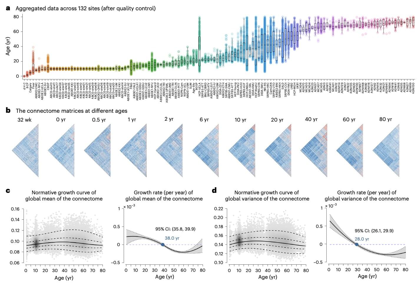
“Functional connectivity of the changes through life,” wrote Lianglong Sun, Tengda Zhao and their colleagues in their paper. “We assemble task-free functional and structural magnetic resonance imaging data from 33,250 individuals at 32 weeks of postmenstrual age to 80 years from 132 global sites.”

Many behavioral studies suggest that using landmarks to navigate through large-scale spaces—known as map-based navigation—is not established until around age 12.

A neuroscience study at Emory University counters that assumption. Through experiments combining and a the researchers dubbed Tiny Town, they showed that five-year-olds have a brain system that supports map-based navigation.

The journal Proceedings of the National Academy of Sciences has published the finding, the first neural evidence that this cognitive ability is in place in such young children.

Mind blanking, when the mind temporarily goes blank, is a distinct, common mental state influenced by attention, arousal, and brain activity patterns. Mind blanking is a common but poorly defined mental phenomenon, encompassing experiences that range from mild drowsiness to a complete loss of con

“Theories are like toothbrushes,” it’s sometimes said. “Everybody has their own and nobody wants to use anybody else’s.”

It’s a joke, but when it comes to the study of consciousness – the question of how we have a subjective experience of anything at all – it’s not too far from the truth.

In 2022, British neuroscientist Anil Seth and I published a review listing 22 theories based in the biology of the brain. In 2024, operating with a less restrictive scope, US public intellectual Robert Kuhn counted more than 200.

A decade ago, University at Buffalo researchers shed some light on an enduring neuroscience mystery: How exactly does a mutated huntingtin protein (HTT) cause Huntington’s disease?

They found that HTT is something like a traffic controller inside neurons, moving different cargo along neuronal highways called axons in concert with other proteins that are key for cellular function and survival. Reduce the amount of non-mutant HTT and you’ll create the neurological equivalent of traffic jams and roadblocks.

Now, the researchers have learned more about what can control the traffic-controlling HTT.

Individuals with retinal degenerative diseases struggle to restore vision due to the inability to regenerate retinal cells. Unlike cold-blooded vertebrates, mammals lack Müller glia (MG)-mediated retinal regeneration, indicating the limited regenerative capacity of mammalian MG. Here, we identify prospero-related homeobox 1 (Prox1) as a key factor restricting this process. Prox1 accumulates in MG of degenerating human and mouse retinas but not in regenerating zebrafish. In mice, Prox1 in MG originates from neighboring retinal neurons via intercellular transfer. Blocking this transfer enables MG reprogramming into retinal progenitor cells in injured mouse retinas. Moreover, adeno-associated viral delivery of an anti-Prox1 antibody, which sequesters extracellular Prox1, promotes retinal neuron regeneration and delays vision loss in a retinitis pigmentosa model. These findings establish Prox1 as a barrier to MG-mediated regeneration and highlight anti-Prox1 therapy as a promising strategy for restoring retinal regeneration in mammals.


Recovery for mammalian retinal degeneration is limited by a lack of Müller glia (MG)-mediated regeneration. Here authors show blocking Prox1 accumulation and intercellular transfer from retinal neurons enables MG reprogramming of retinal progenitor cells, promotes retinal neuron regeneration, and delays vision loss.