Functional unblinding was common in most psychedelic randomized clinical trials for psychiatric disorders, with 70% correctly identifying treatment allocation, raising concerns for trial validity.
Question What is the prevalence of blinding integrity assessment and the extent of functional unblinding in psychedelic randomized clinical trials (RCTs) for psychiatric disorders?
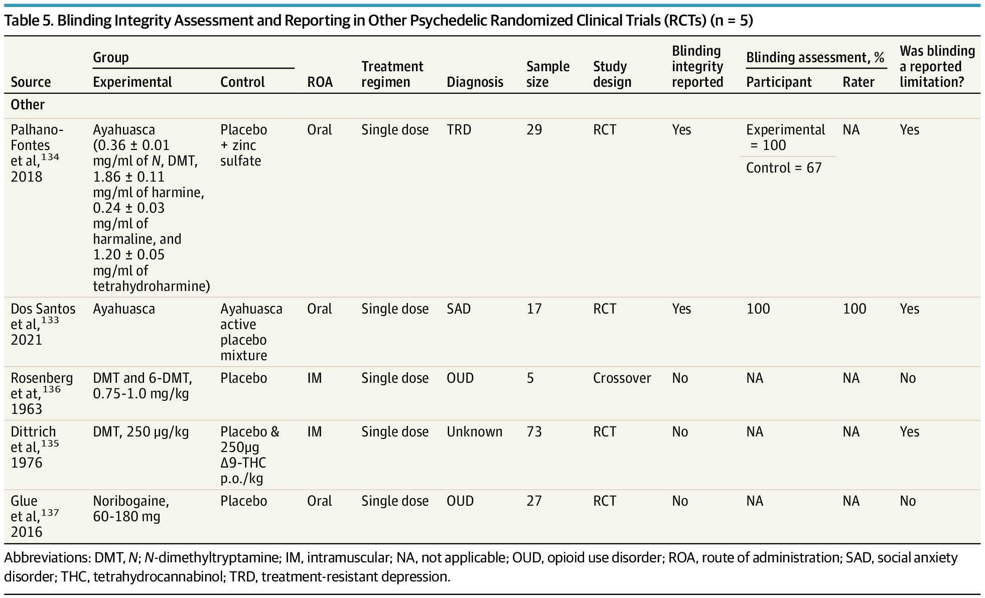
Findings Of 112 RCTs identified, 29.5% (n = 33) evaluated blinding integrity. Functional unblinding was substantial: psilocybin, lysergic acid diethylamide (LSD), and ayahuasca studies frequently reported blinding failure values of more than 90% among participants and raters; inert placebo-controlled 3,4-methylenedioxymethamphetamine (MDMA) trials exceeded 85%; ketamine trials rarely assessed blinding (17.9%) but showed improved preservation with midazolam vs saline controls.
Meaning Functional unblinding is pervasive in psychedelic RCTs, underscoring the need for standardized assessment methods and improved trial designs to ensure valid efficacy evaluations.