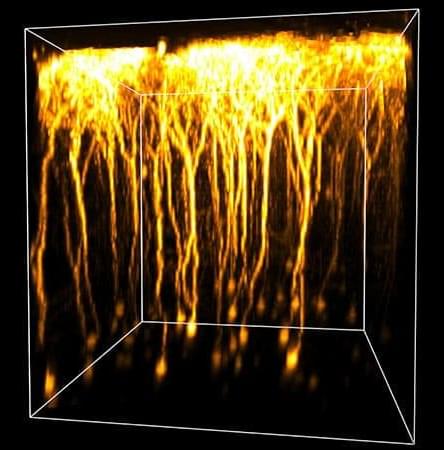
Researchers used a blood protein wash to render brain tissue transparent down to 700 µm, witnessing live-firing activity without killing a single neuron.
Category: neuroscience – Page 38
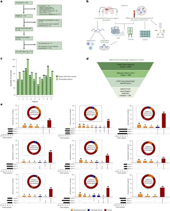
Adjuvant personalized multivalent neoantigen DNA vaccination for MGMT unmethylated glioblastoma: a phase 1 trial
A personalized vaccine to treat glioblastoma, a fast-growing and incurable brain cancer that affects four in 100,000 people in the U.S., is safe and elicits robust and broad immune responses that appears to increase recurrence-free survival in a subset of patients after surgery, according to an early-stage clinical trial co-led by researchers at Washington University School of Medicine in St. Louis.
In patients with an especially aggressive form of glioblastoma, the vaccine caused no serious side effects and prolonged patients’ overall survival compared to historical outcomes after standard-of-care surgery and chemo-radiotherapy. One long-term survivor remains recurrence-free nearly five years later.
The results of the phase 1 trial, conducted at Siteman Cancer Center, based at Barnes-Jewish Hospital and WashU Medicine, were published May 12 in Nature Cancer. The study was led jointly by Mass General Brigham and Geneos Therapeutics, a Philadelphia-based biotechnology company.
“We are extremely encouraged by these results,” said Tanner M. Johanns, MD, PhD, lead author of the study and an assistant professor in the Division of Oncology in the John T. Milliken Department of Medicine at WashU Medicine. “This kind of vaccine is a first for glioblastoma, and it is exciting to think how we can leverage this individualized therapeutic DNA cancer vaccine platform to make a positive impact on the lives of patients who are fighting this disease. Additionally, combination therapies leveraging this personalized platform are currently being investigated at WashU to test if outcomes may be improved further.”
Abstract: Nature Cancer
Johanns and colleagues report the results (including safety, efficacy and immunogenicity) of a phase 1 clinical trial of a DNA-based personalized therapeutic cancer vaccine administered following surgical resection and radiation in patients with MGMT unmethylated glioblastoma.
Hippocampal ripples and replay reveal how brain recombines past knowledge for flexible planning
When facing new situations or problems, humans typically rely on knowledge they acquired in the past. Specifically, neuroscience studies suggest that the brain reorganizes past experiences and previously acquired knowledge, creating mental frameworks that can help humans to solve the problems they are facing. The recombination of past knowledge into new mental structures also allows humans to flexibly plan future actions in changing environments. Past studies suggest that two key brain regions contribute to this process, the hippocampus and the medial prefrontal cortex (mPFC).
The hippocampus is a brain structure that plays a key role in the formation of memories and spatial navigation. The mPFC, on the other hand, is known to support decision-making, planning, reasoning and the integration of information.
Researchers at Beijing Normal University, the Chinese Academy of Medical Sciences, University College London (UCL) and other institutes recently set out to investigate how the hippocampus and mPFC work together to combine past knowledge into new configurations. Their findings, published in Nature Neuroscience, suggest that this process is supported by brief bursts of high-frequency neural activity in the hippocampus, called hippocampal ripples, and the replay (i.e., re-activation) of past experiences in the brain.
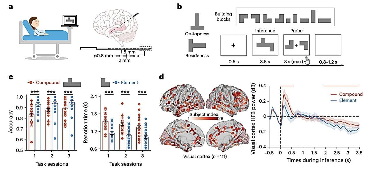
Decoding intended speech with an intracortical brain-computer interface in a person with long-standing anarthria and locked-in syndrome
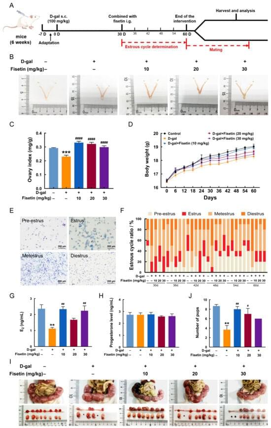
This study aimed to explore the alleviating effects of fisetin, a polyphenolic flavonoid, on ovarian dysfunction in a D-galactose (D-gal)-induced aging mouse model, as well as the underlying mechanisms, using both in vivo and in vitro experiments. Mice were subcutaneously injected with D-gal (100 mg/kg/day) for 60 days to establish the ovarian aging model; during the final 30 days, fisetin (10, 20, 30 mg/kg/day) was given orally. In addition, a senescent model of granulosa cell (GC) was established using D-gal and treated with fisetin. Fisetin supplementation improved ovarian endocrine function and reproductive capacity in aging mice, as reflected by regularized estrous cycles, elevated estradiol levels, and increased embryo numbers. Furthermore, fisetin reduced the number of atretic follicles and the extent of ovarian fibrosis and senescence, while simultaneously restoring the proliferation-apoptosis balance in follicular GCs, as well as alleviating oxidative stress. RNA-sequencing revealed that AMP-activated protein kinase (AMPK)/mechanistic target of rapamycin (mTOR) signaling and mitophagy were involved in the protective effects of fisetin against ovarian aging. Consistently, fisetin treatment promoted mitophagy, accompanied by AMPK/mTOR activation in ovarian tissues and GCs following D-gal exposure. Inhibition of AMPK attenuated the effect of fisetin on mitophagy. Additionally, blockage of mitophagy also reversed the beneficial effects of fisetin on mitochondrial injury, oxidative stress, cell cycle arrest, and cellular senescence in D-gal-induced senescent GCs. These findings indicate that fisetin prevents ovarian aging by suppressing follicular GC oxidative damage and ameliorating cell cycle arrest via activation of AMPK/mTOR-mediated mitophagy, thereby preserving female fertility.

Sleep Duration Linked to Accelerated Aging
The relationship between sleep and disease suggests that there exists a connection between the brain and the body that extends beyond merely influencing the brain itself.
Among brain-related disorders, short sleep was significantly associated with depressive episodes and anxiety disorders, as seen in other studies of sleep and mental health. Short sleep was also associated with obesity, type 2 diabetes, hypertension, ischemic heart disease, and heart arrhythmias.
Short and long sleep were associated with chronic obstructive pulmonary disease, asthma, and a cluster of digestive disorders, including gastritis and gastroesophageal reflux disease.
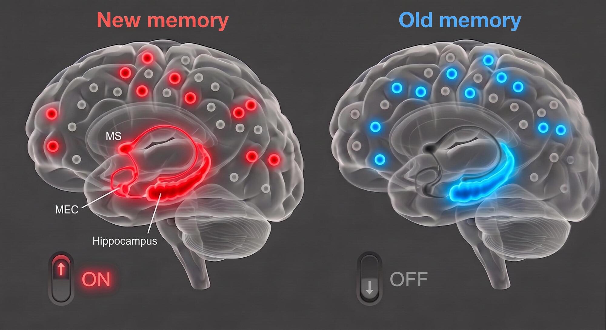
How the brain switches between older and newer memories
As humans and other animals experience new things, their brains continuously update their memory of past events. These updates allow them to adapt to changing environments, all while preserving older memories that could still help them to make decisions in some situations.
Many past neuroscience studies have investigated the neural circuits involved in the encoding and retrieval of memories. However, the mechanisms via which it decides whether to retrieve older or newly updated memories remain poorly understood.
Researchers at Korea Advanced Institute of Science and Technology (KAIST) recently carried out a study involving mice that was aimed at better understanding how the brain switches between older and newer memories.
This Physicist (Unexpectedly) Derived Gravity from Information
Sponsors: Accelerate your efficiency. Sign up for your one-dollar-per-month trial today at http://shopify.com/theories Sign up for Claude today at http://claude.ai/theoriesofeverything and checkout Claude Pro — which includes access to all of the features mentioned in today’s episode. I personally subscribe to The Economist. FLASH SALE to May 18: 50% off annual (double the usual!) No other podcast has this! https://economist.com/TOE
What if gravity is just entropy in disguise? Professor Erik Verlinde joins me to argue that gravity isn’t a fundamental force—it’s thermodynamic, emerging from quantum information the way gas pressure emerges from molecules bouncing around. We explore why spacetime may be stitched together by entanglement, and how dark energy and dark matter both pop out automatically without extra particles or parameters. Verlinde explains why the cosmological constant problem is a red herring, and why there may be no final theory of physics. When asked where the universe comes from, his answer is one word: chaos.
SUPPORT: Support me on Substack: https://curtjaimungal.substack.com/su… me on Crypto: https://commerce.coinbase.com/checkou… Support me on PayPal: https://www.paypal.com/donate?hosted_… JOIN MY SUBSTACK (Personal Writings): https://curtjaimungal.substack.com LISTEN ON SPOTIFY: https://open.spotify.com/show/4gL14b9… TIMESTAMPS:
- 00:00:00 — Thermodynamic Gravity and Information
- 00:06:35 — Beyond Effective Field Theory
- 00:13:08 — Turtles All The Way Down
- 00:25:41 — Entropy as a Force
- 00:36:31 — Entanglement and Spatial Connectivity
- 00:47:31 — Deriving Inertia and F=ma
- 00:56:41 — De Sitter Space Challenges
- 01:02:01 — Dark Matter and Milgram
- 01:11:51 — The Emergence of Time
- 01:21:01 — Statistical Gravity Fluctuations
- 01:27:01 — Quantum Computational Complexity
- 01:36:01 — Physics Intuition and Mentorship
- 01:47:31 — Beauty, Garbage, and Chaos
LINKS MENTIONED: Papers, books, websites:
- https://scholar.google.com/citations?…
- https://journals.aps.org/prb/pdf/10.1…
- https://arxiv.org/abs/1001.0785
- https://arxiv.org/abs/1611.02269
- https://journals.aps.org/pr/pdf/10.11…
- https://amazon.com/dp/0486600688?tag=…
- https://www.nature.com/articles/248030a0
- https://en.wikisource.org/wiki/Transl…
- https://arxiv.org/abs/gr-qc/9504004
- https://arxiv.org/abs/hep-th/0603001
- https://arxiv.org/abs/1005.3035
- https://arxiv.org/abs/hep-th/0106112
- https://arxiv.org/pdf/1408.3203
- https://arxiv.org/abs/1911.02087
- https://arxiv.org/abs/1905.08255
- https://www.ias.edu/sns/events/iaspct…
- https://amazon.com/dp/0262533413?tag=…
Videos:
-
• A 2 Hour Deep Dive into Entropy
-
• The Mathematics of String Theory [Graduate…
-
• The Debate That Divides Physics: Is the Un…
-
• The Physicist Who Found Quantum Theory’s U…
-
• Retrocausality & The Transactional Interpr…
-
• The Physicist Who Proved Entropy = Gravity
-
• The Physicist Who Says Time Doesn’t Exist
-
• The Most Astonishing Theory of Black Holes
-
• The (Simple) Theory That Explains Everythi…
-
• The Crisis in String Theory is Worse Than…
-
• Dark Dimensions: NEW THEORY Unifying Dark…
-
• MIT Scientist’s Discovery: “Black Holes Mi…
-
• The Woman Who Broke Gravity | Claudia de Rham
-
• Solving the Problem of Consciousness | Ste…
-
• Frederic Schuller: The Physicist Who Deriv…
-
• The Loop Quantum Gravity Debacle: Carlo Ro…
-
• An (Elementary) Introduction to Quantum Co…
-
• Can Physics Explain Its Own Laws?
-
• The Nobel Laureate Who (Also) Says Quantum…
-
• This Cosmologist Discovered Something Stra…
-
• Michael Levin: Consciousness, Biology, Uni…
SOCIALS:
- Twitter:
/ toewithcurt
- Discord Invite:
/ discord
Guests do not pay to appear. Theories of Everything receives revenue solely from viewer donations, platform ads, and clearly labelled sponsors; no guest or associated entity has ever given compensation, directly or through intermediaries. #science.
JOIN MY SUBSTACK (Personal Writings): https://curtjaimungal.substack.com.
LISTEN ON SPOTIFY: https://open.spotify.com/show/4gL14b9…
TIMESTAMPS: 00:00:00 — Thermodynamic Gravity and Information 00:06:35 — Beyond Effective Field Theory 00:13:08 — Turtles All The Way Down 00:25:41 — Entropy as a Force 00:36:31 — Entanglement and Spatial Connectivity 00:47:31 — Deriving Inertia and F=ma 00:56:41 — De Sitter Space Challenges 01:02:01 — Dark Matter and Milgram 01:11:51 — The Emergence of Time 01:21:01 — Statistical Gravity Fluctuations 01:27:01 — Quantum Computational Complexity 01:36:01 — Physics Intuition and Mentorship 01:47:31 — Beauty, Garbage, and Chaos.