Though we learn so much during our first years of life, we can’t, as adults, remember specific events from that time. Researchers have long believed we don’t hold onto these experiences because the part of the brain responsible for saving memories — the hippocampus — is still developing well into adolescence and just can’t encode memories in our earliest years. But new Yale research finds evidence that’s not the case.
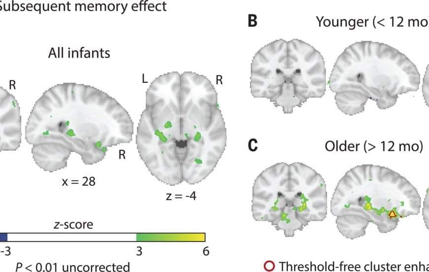
In a study, the researchers showed infants new images and later tested whether they remembered them. When an infant’s hippocampus was more active upon seeing an image the first time, they were more likely to appear to recognize that image later.
The findings, published in Science, indicate that memories can indeed be encoded in our brains in our first years of life. And the researchers are now looking into what happens to those memories over time.