Before you even know what a word means, your brain is already playing a rapid-fire game of linguistic LEGO. Discover how our minds secretly dissect words, piece by orthographic piece, in the blink of an eye.
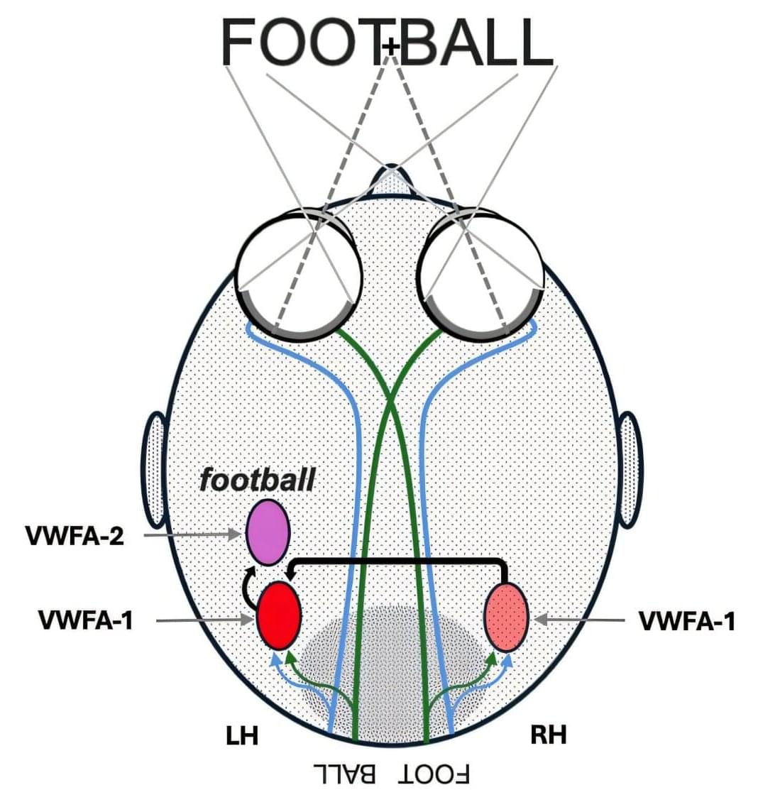
Imagine catching a flash of the word football on a screen. Before you even register its meaning (“a game” or “a ball”), your brain may have already parsed it into “foot” + “ball.” A clever new experiment used red-and-blue anaglyph glasses and split-second word flashes to probe this. It found that real compound words (like football) are recognized much faster than lookalikes (like shamrock), suggesting our eyes and brain latch onto word form almost instantly.
In the lab, volunteers wore 3D-style red/blue glasses while words appeared for just 60 milliseconds under a mask. Each word was painted half red and half blue, splitting it either at a meaningful break or in the middle of a syllable. For example, “FOOT” might be blue and “BALL” red, or vice versa, sending “foot” to one hemisphere and “ball” to the other. Participants then quickly reported if what they saw was a real word or a made-up one.