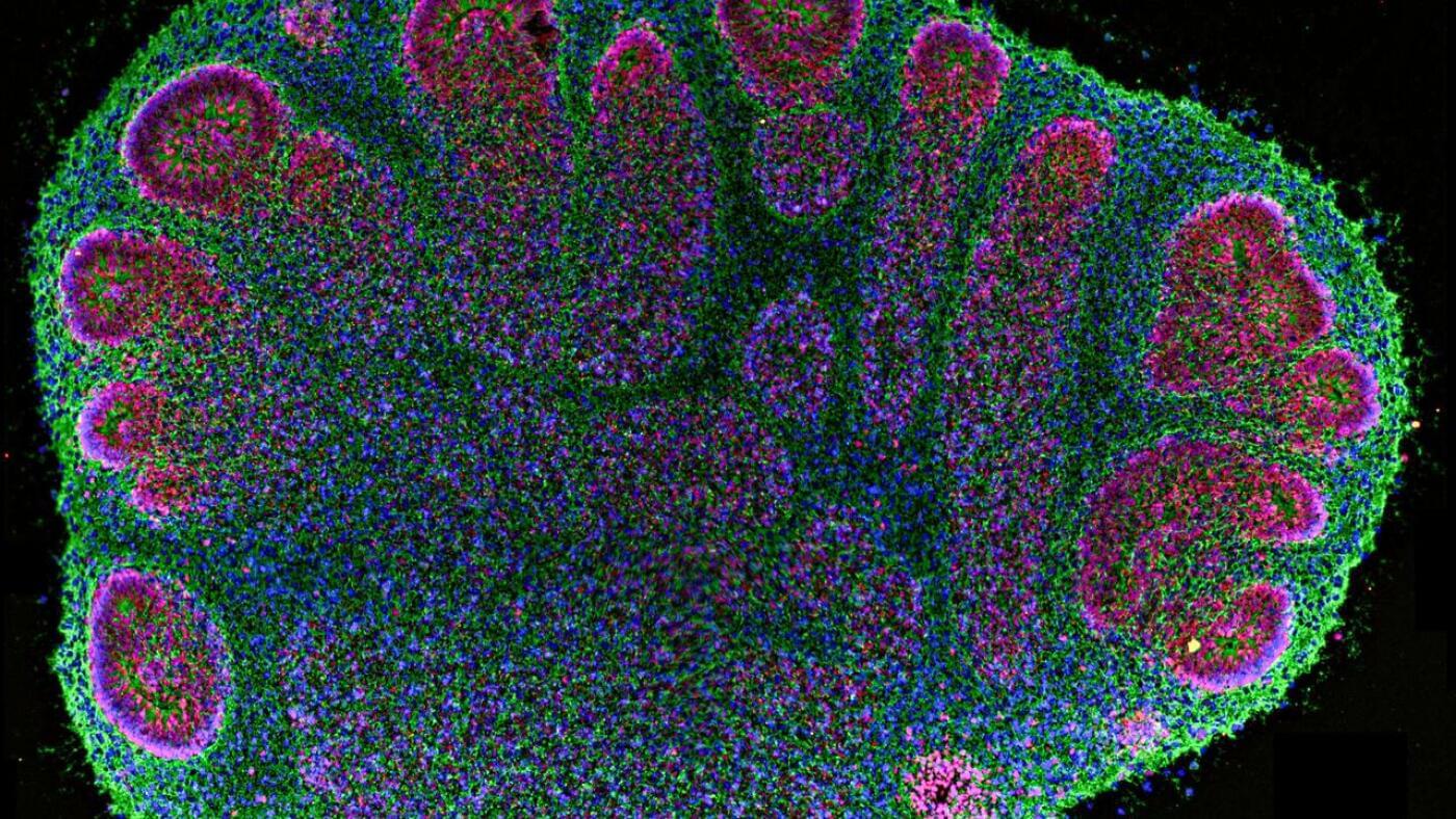
Pea-size clusters of human cells called brain organoids inspire both hope and fear. Experts are debating how scientists can responsibly use these bits of gray matter.

The secret to a healthier and “younger” heart lies in the vagus nerve. A recent study published in Science Translational Medicine has shown that preserving bilateral cardiac vagal innervation is an anti-aging factor. In particular, the right cardiac vagus nerve emerges as a true guardian of cardiomyocyte health, helping to preserve the longevity of the heart independently of heart rate.
‘When the integrity of the connection to the vagus nerve is lost, the heart ages more rapidly,’ explains the senior author.
‘Even partial restoration of the connection between the right vagus nerve and the heart is sufficient to counteract the mechanisms of remodelling and preserve effective cardiac contractility,’ adds another author.
‘We have developed an implantable bioabsorbable nerve conduit designed to promote and guide the spontaneous regeneration of the thoracic vagus nerve at the cardiac level,’ explains a co-author.
Treated adult male minipigs displayed improved global circumferential, longitudinal, and radial strains and reduced diastolic dyssynchrony. Histological analysis revealed partial repair with about 20% viable vagal fascicles, restoration of myocardial parasympathetic fibers, normalization of oxidative stress and aging markers, and prevention of interstitial fibrosis.
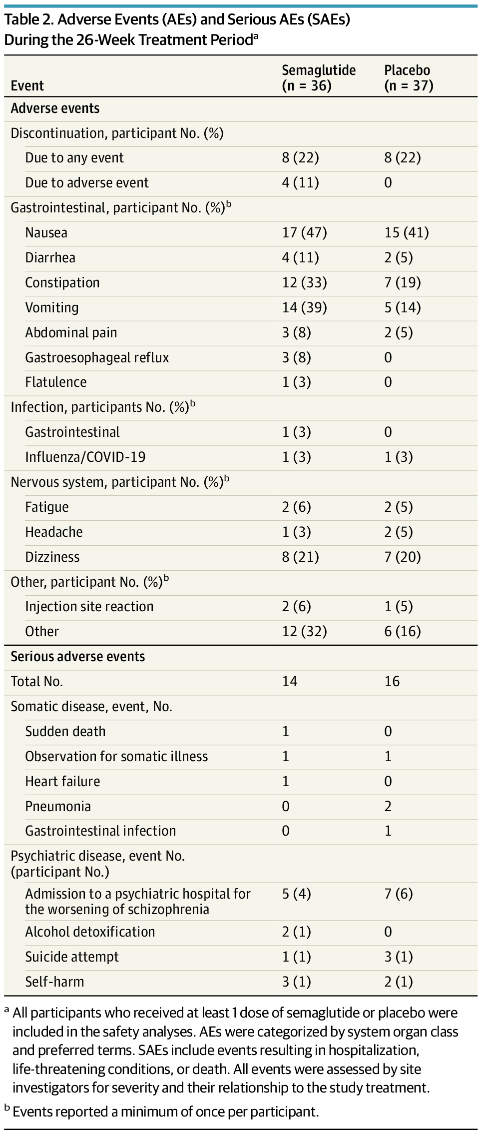
Semaglutide reduced HbA1c and body weight over 26 weeks in individuals with schizophrenia spectrum disorders and early glycemic abnormalities taking clozapine or olanzapine.
Question Can adjunctive semaglutide improve glycemic control and weight outcomes in individuals with early-stage prediabetes or diabetes and schizophrenia spectrum disorders who initiated clozapine or olanzapine within the past 5 years?
Findings In this randomized clinical trial including 73 participants, semaglutide was found to significantly reduce hemoglobin A1c level and body weight over a 26-week period. Approximately one-half of individuals treated with semaglutide achieved low-risk hemoglobin A1c levels, compared with the placebo group.
Meaning This study found that semaglutide can mitigate the early metabolic burden associated with second-generation antipsychotic use in schizophrenia and may support the prevention of long-term cardiometabolic complications when initiated during the early stages of metabolic dysregulation.
A study co-led by Richard Carson and colleagues at the Yale School of Medicine has identified a measurable molecular difference in the brains of autistic adults – finding reduced availability of a key glutamate receptor involved in brain signaling balance.
Published in The American Journal of Psychiatry, the research offers new insight into the biological mechanisms of autism and could inform future diagnostic tools and targeted supports.
👉 Read the full story.
Brains of autistic individuals have fewer of a specific kind of glutamate receptor, supporting an idea that autism is driven by a signaling imbalance.
Humans have the remarkable ability to remember the same person or object in completely different situations. We can easily distinguish between dinner with a friend and a business meeting with the same friend. “We already know that deep in the memory centers of the brain, specific cells, called concept neurons, respond to this friend, regardless of the environment in which he appears,” says Prof. Florian Mormann from the Clinic for Epileptology at the UKB, who is also a member of the Transdisciplinary Research Area (TRA) Life & Health at the University of Bonn.
However, the brain must be able to combine this content with the context in order to form a useful memory. In rodents, individual neurons often mix these two pieces of information. “We asked ourselves: Does the human brain function fundamentally differently here? Does it map content and context separately to enable a more flexible memory? And how do these separate pieces of information connect when we need to remember specific content according to context?” says Dr. Marcel Bausch, working group leader at the Department of Epileptology and member of TRA Life & Health at the University of Bonn.
Past research has found that exposure to bright lights and high levels of noise can alter both physiological processes and human behavior. For instance, an elevated or limited exposure to bright lights and noise has been found to influence people’s sleeping patterns, circadian rhythm, mood, metabolism, stress levels and mental performance.
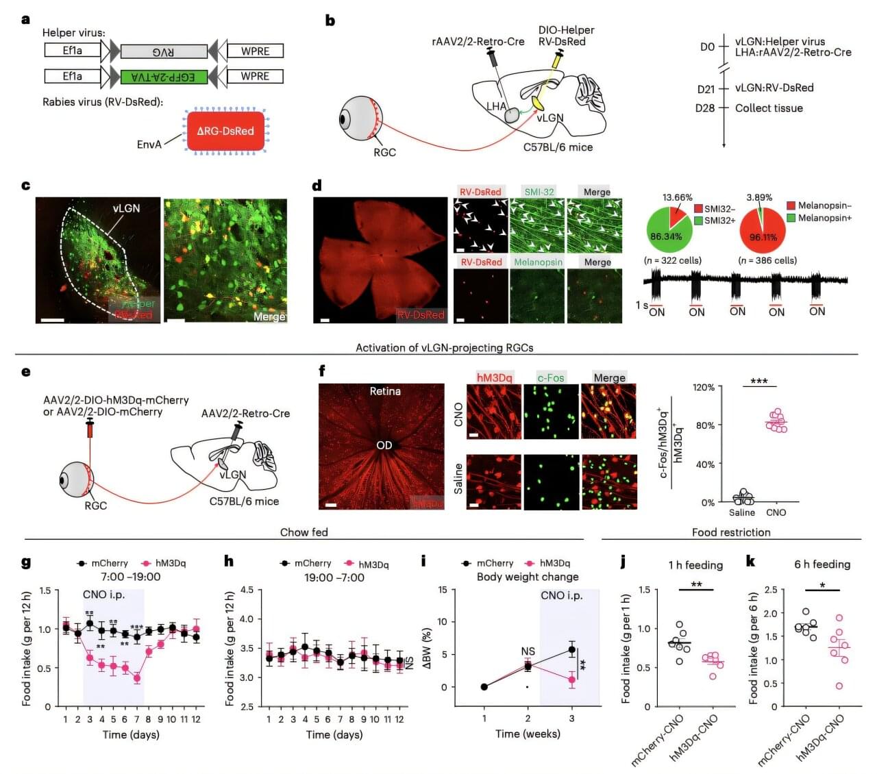
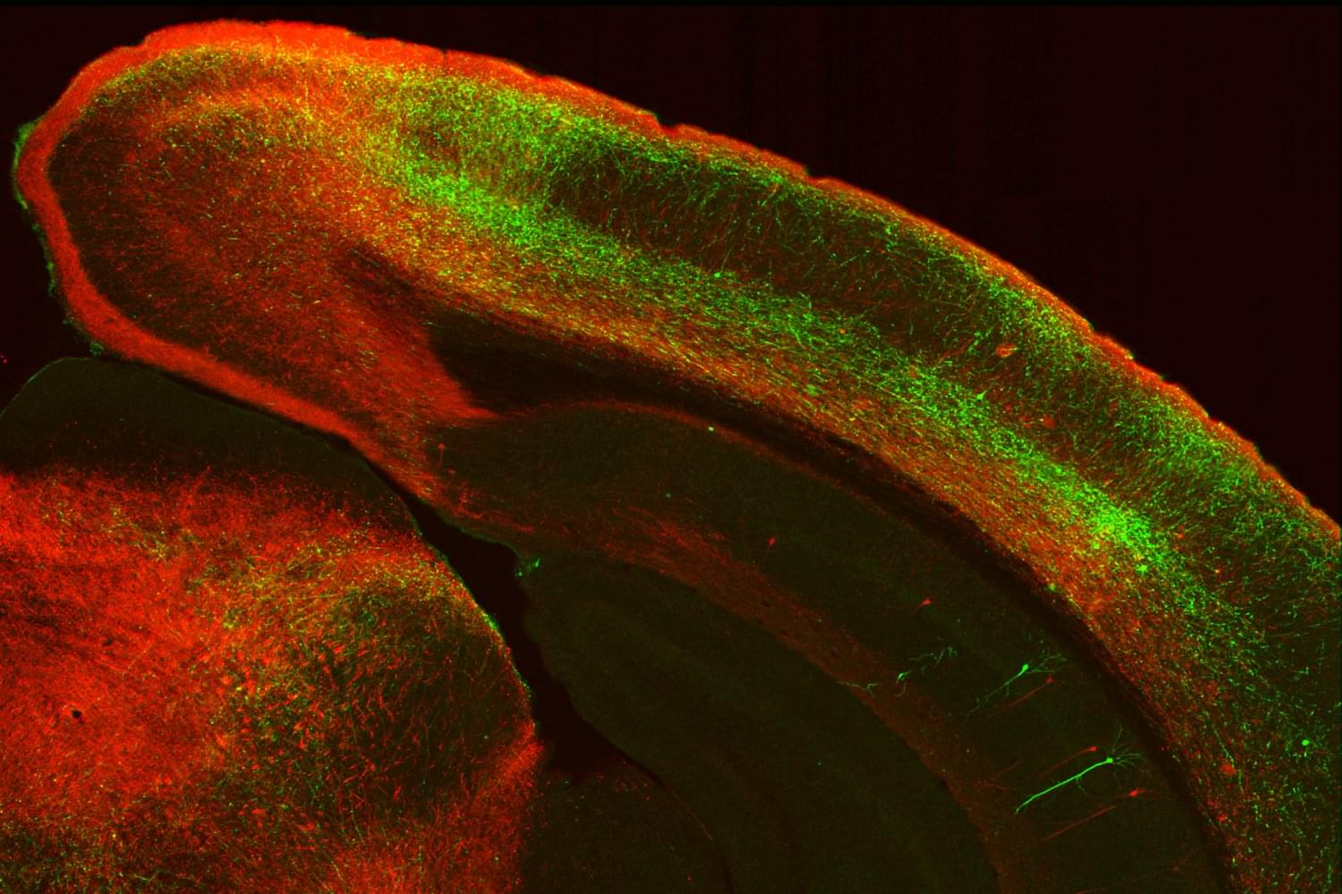
Researchers at Jinan University and other institutes in China recently carried out a new study involving mice, exploring the possibility that the exposure to bright lights also influences eating behavior and body weight. Their findings, published in Nature Neuroscience, suggest that bright light exposure suppresses food consumption in mice and can lead to weight loss, while also identifying neural processes that could support these light-induced changes in feeding behavior.
“Environmental light regulates nonimage-forming functions like feeding, and bright light therapy shows anti-obesity potential, yet its neural basis remains unclear,” wrote Wen Li, Xiaodan Huang and their colleagues in their paper. “We show that bright light treatment effectively reduces food intake and mitigates weight gain in mice through a visual circuit involving the lateral hypothalamic area (LHA).”

For more than a century, scientists have wondered why physical structures like blood vessels, neurons, tree branches, and other biological networks look the way they do. The prevailing theory held that nature simply builds these systems as efficiently as possible, minimizing the amount of material needed. But in the past, when researchers tested these networks against traditional mathematical optimization theories, the predictions consistently fell short.
The problem, it turns out, was that scientists were thinking in one dimension when they should have been thinking in three. “We were treating these structures like wire diagrams,” Rensselaer Polytechnic Institute (RPI) physicist Xiangyi Meng, Ph.D., explains. “But they’re not thin wires, they’re three-dimensional physical objects with surfaces that must connect smoothly.”
This month, Meng and colleagues published a paper in the journal Nature showing that physical networks in living systems follow rules borrowed from an unlikely source: string theory, the exotic branch of physics that attempts to explain the fundamental structure of the universe.
Pain-sensing neurons in the gut kindle inflammatory immune responses that cause allergies and asthma, according to a new study by Weill Cornell Medicine. The findings, published in Nature, suggest that current drugs may not be as effective because they only address the immune component of these conditions, overlooking the contribution of neurons.
“Today’s blockbuster biologics are sometimes only 50% effective and when the treatments do work, they sometimes lose their efficacy over time,” said senior author Dr. David Artis, director of the Jill Roberts Institute for Research in Inflammatory Bowel Disease and the Michael Kors Professor in Immunology at Weill Cornell.
While the idea may be new to the field, Dr. Artis has been thinking about the role the nervous system may play in allergies and asthma for about two decades. For example, many of the symptoms that characterize these conditions, like itching and wheezing, are known to be neuronally controlled. “That was one of the clues that prompted us to look closer for a connection,” Dr. Artis said.