Toggle light / dark theme

The Royal Swedish Academy of Sciences has decided to award the Nobel Prize in Chemistry 2017 to Jacques Dubochet (University of Lausanne, Switzerland), Joachim Frank (Columbia University, New York, USA) and Richard Henderson (MRC Laboratory of Molecular Biology, Cambridge, UK) “for developing cryo-electron microscopy for the high-resolution structure determination of biomolecules in solution”.

We may soon have detailed images of life’s complex machineries in atomic resolution. The Nobel Prize in Chemistry 2017 is awarded to Jacques Dubochet, Joachim Frank and Richard Henderson for the development of cryo-electron microscopy, which both simplifies and improves the imaging of biomolecules. This method has moved biochemistry into a new era.

A picture is a key to understanding. Scientific breakthroughs often build upon the successful visualisation of objects invisible to the human eye. However, biochemical maps have long been filled with blank spaces because the available technology has had difficulty generating images of much of life’s molecular machinery. Cryo-electron microscopy changes all of this. Researchers can now freeze biomolecules mid-movement and visualise processes they have never previously seen, which is decisive for both the basic understanding of life’s chemistry and for the development of pharmaceuticals.

Read more

Light-activated nanoparticles, also known as quantum dots, can provide a crucial boost in effectiveness for antibiotic treatments used to combat drug-resistant superbugs such as E. coli and Salmonella, new University of Colorado Boulder research shows.

Multi-drug resistant pathogens, which evolve their defenses faster than new can be developed to treat them, cost the United States an estimated $20 billion in direct healthcare costs and an additional $35 billion in lost productivity in 2013.

CU Boulder researchers, however, were able to re-potentiate existing for certain clinical isolate infections by introducing nano-engineered dots, which can be deployed selectively and activated or de-activated using specific wavelengths of light.

Read more

(Phys.org)—A team of researchers from the University of California and the University of Tokyo has found a way to use the CRISPR gene editing technique that does not rely on a virus for delivery. In their paper published in the journal Nature Biomedical Engineering, the group describes the new technique, how well it works and improvements that need to be made to make it a viable gene editing tool.

CRISPR-Cas9 has been in the news a lot lately because it allows researchers to directly edit genes—either disabling unwanted parts or replacing them altogether. But despite many success stories, the technique still suffers from a major deficit that prevents it from being used as a true medical tool—it sometimes makes mistakes. Those mistakes can cause small or big problems for a host depending on what goes wrong. Prior research has suggested that the majority of mistakes are due to delivery problems, which means that a replacement for the virus part of the technique is required. In this new effort, the researchers report that they have discovered just a such a replacement, and it worked so well that it was able to repair a in a Duchenne muscular dystrophy mouse model. The team has named the CRISPR-Gold, because a gold nanoparticle was used to deliver the molecules instead of a virus.

The new package was created by modifying a bit of DNA to cause it to stick to a gold nanoparticle and then a Cas9 protein and also an RNA guide. The package was then coated with a polymer that served as a containment casing—one that also triggered endocytosis (a form of cell transport) and helped the molecules escape endosomes once inside the target cells. The molecules then set to work—the Cas9 cut the target DNA strand, the guide RNA showed what needed to be done and a DNA strand was placed where a mutation had existed. The result was a gene free of a mutation that causes Duchenne muscular dystrophy.

Read more

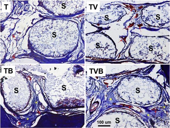
Muscle from a mouse model of Duchenne muscular dystrophy. Fibrous scar tissue is in blue and healthy muscle is in pink. CONBOY LAB AND MURTHY LAB While promising, applications of CRISPR-Cas9 gene editing have so far been limited by the challenges of delivery—namely, how to get all the CRISPR parts to every cell that needs them. In a study published today (October 2) in Nature Biomedical Engineering, researchers have successfully repaired a mutation in the gene for dystrophin in a mouse model of Duchenne muscular dystrophy by injecting a vehicle they call CRISPR-Gold, which contains the Cas9 protein, guide RNA, and donor DNA, all wrapped around a tiny gold ball.

The authors have made “great progress in the gene editing area,” says Tufts University biomedical engineer Qiaobing Xu, who did not participate in the work but penned an accompanying commentary. Because their approach is nonviral, Xu explains, it will minimize the potential off-target effects that result from constant Cas9 activity, which occurs when users deliver the Cas9 template with a viral vector.

Duchenne muscular dystrophy is a degenerative disease of the muscles caused by a lack of the protein dystrophin. In about a third of patients, the gene for dystrophin has small deletions or single base mutations that render it nonfunctional, which makes this gene an excellent candidate for gene editing. Researchers have previously used viral delivery of CRISPR-Cas9 components to delete the mutated exon and achieve clinical improvements in mouse models of the disease.

Read more

Scientists at The University of Manchester have created the world’s first ‘molecular robot’ that is capable of performing basic tasks including building other molecules. The tiny robots, which are a millionth of a millimetre in size, can be programmed to move and build molecular cargo, using a tiny robotic arm.

Each individual robot is capable of manipulating a single molecule and is made up of just 150 carbon, hydrogen, oxygen and nitrogen atoms. To put that size into context, a billion billion of these robots piled on top of each other would still only be the same size as a single grain of salt. The robots operate by carrying out chemical reactions in special solutions which can then be controlled and programmed by scientists to perform the basic tasks.

In the future such robots could be used for medical purposes, advanced manufacturing processes and even building molecular factories and assembly lines. The research will be published in Nature on Thursday 21st September.

Read more

Summary: Nanodocs? #Swallow #the #doctor? The authors of a recent research study, says soon we will be able to “swallow the surgeon.” Using medical #nanobots to diagnose and treat disease from inside the body. Study authors documented recent advances in nanotechnology tools, such as nanodrillers, microgrippers, and microbullets – and show how #nanodocs have tremendous potential in the areas of precision surgery, detection, detoxification and targeted drug delivery.


Summary: Nanodocs? Swallow the doctor? The authors of a recent research study, say the concept of “swallow the surgeon” – or using medical nanobots to diagnose and treat disease from inside the body – may be closer than we think. Study authors document recent advances in nanotechnology tools, such as nanodrillers, microgrippers, and microbullets – and show how nanodocs have tremendous potential in the areas of precision surgery, detection, detoxification and targeted drug delivery. Cover photo: The old way to swallow the surgeon. Credit: R. Collin Johnson / Attributed to Stanford University.

Imagine that you need to repair a defective heart valve, a major surgery. Instead of ripping your chest cut open, a doctor merely injects you with a syringe full of medical nanorobots, called nanodocs for short. You emerge from the ‘surgery’ unscathed, and your only external wound is the puncture hole from the injection.

According to a recent study published by nanorobotic engineers at the University of California San Diego (UCSD), the concept of ‘swallow the doctor’ may be closer to reality than we think.

Cellulose is the most abundant organic polymer in the world. It is the primary compound in the cell walls of green plants, and is typically used to make paper and cardboard.

At the VTT Technical Centre of Finland, a state owned research and development non-profit, scientists have used nano-structured cellulose to make a 3D printable material.

The nanocellulose paste is now in development to make smart-dressings that heal and monitor skin wounds.

Read more

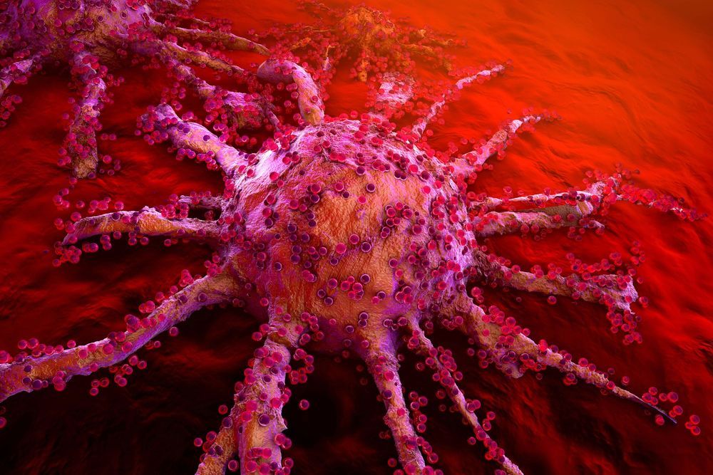
The new self-propelled, cancer-seeking bacteriobot swims right into the tumor and zaps it with a deadly payload of cancer drugs.

The recently perfected #bacteriobot holds ‘a lot of promise’ in treating #cancer says a physician. Cancer patients at a hospital in Montreal may be the first to be treated with these #nanorobots built out of bacteria.


Summary: The recently perfected bacteriobot holds ‘a lot of promise’ in treating cancer says a physician. Cancer patients at a hospital in Montreal may be the first to be treated with nanorobots built out of bacteria. The new self-propelled, cancer-seeking bacteriobot swims right into the tumor and zaps it with a deadly payload of cancer drugs. [Cover image: Getty Images/iStock.]

Google’s Futurist Ray Kurzweil once said that within decades, we will have nanobots, swimming through our veins keeping us healthy. The tiny robots will keep us healthy by correcting DNA errors, removing toxins, extending our memories and zapping cancer. The Futurist said that back in 2007, and his prophecy is becoming a reality, at least in the treatment of cancer.