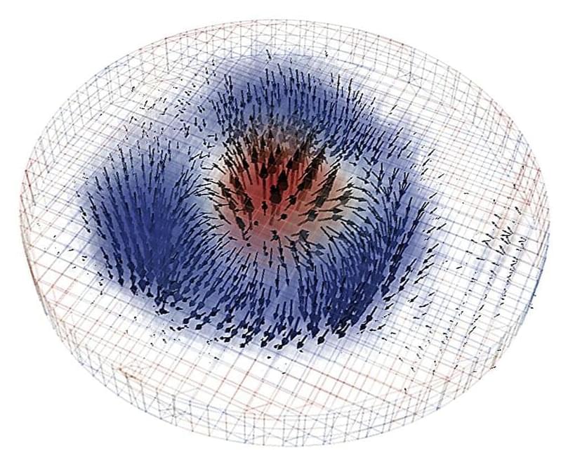
Researchers at Berkeley Lab have advanced the understanding of magnetic skyrmions by developing techniques to image their 3D structures.
These nanoscale objects show promise for revolutionizing microelectronics through enhanced data storage capabilities and reduced energy consumption.
A difficult-to-describe nanoscale structure called the magnetic skyrmion holds potential for creating advanced microelectronic devices, including those with vast data storage capacities and significantly lower power requirements.
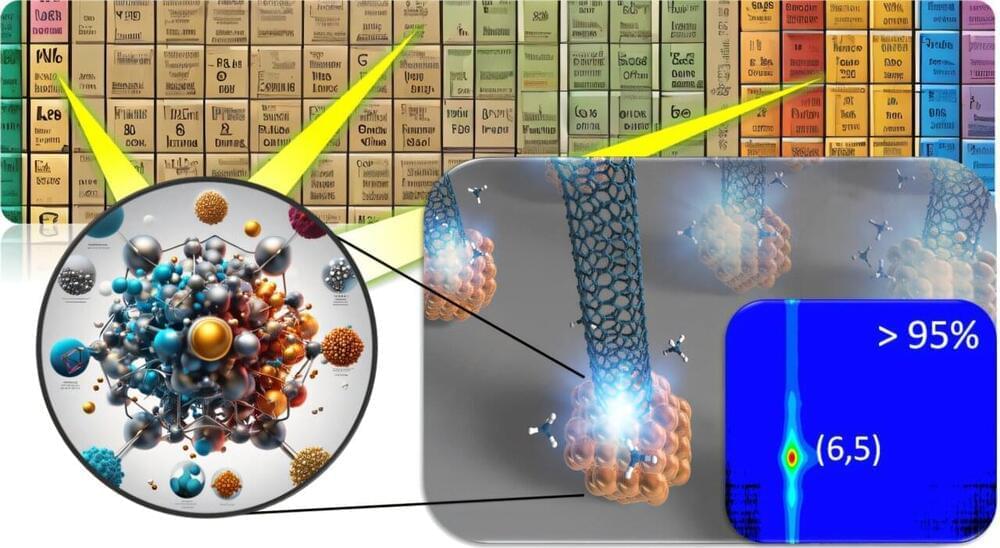
Researchers have achieved a significant breakthrough in the synthesis of carbon nanotubes (CNTs) by developing a novel catalyst that allows for precise control over their atomic arrangement, known as chirality. This advancement paves the way for the creation of innovative semiconductor devices, addressing a challenge that has remained unresolved for over 30 years.
A difficult-to-describe nanoscale object called the magnetic skyrmion might one day yield new microelectronic devices that can do much more—for example, massive data storage—all while consuming much less power.
The great George Church takes us through the revolutionary journey of DNA sequencing from his early groundbreaking work to the latest advancements. He discusses the evolution of sequencing methods, including molecular multiplexing, and their implications for understanding and combating aging.
We talk about the rise of biotech startups, potential future directions in genome sequencing, the role of precise gene therapies, the ongoing integration of nanotechnology and biology, the potential of biological engineering in accelerating evolution, transhumanism, the Human Genome Project, and the importance of intellectual property in biotechnology.
The episode concludes with reflections on future technologies, the importance of academia in fostering innovation, and the need for scalable developments in biotech.
00:00 Introduction to Longevity and DNA Sequencing. 01:43 George Church’s Early Work in Genomic Sequencing. 02:38 Innovations in DNA Sequencing. 03:15 The Evolution of Sequencing Methods. 07:41 Longevity and Aging Reversal. 12:12 Biotech Startups and Commercial Endeavors. 17:38 Future Directions in Genome Sequencing. 28:10 Humanity’s Role and Transhumanism. 37:23 Exploring the Connectome and Neural Networks. 38:29 The Mystery of Life: From Atoms to Living Systems. 39:35 Accelerating Evolution and Biological Engineering. 41:37 Merging Nanotechnology and Biology. 45:00 The Future of Biotech and Young Innovators. 47:16 The Human Genome Project: Successes and Shortcomings. 01:01:10 Intellectual Property in Biotechnology. 01:06:30 Future Technologies and Final Thoughts.
In 2019, researchers from the Massachusetts Institute of Technology made headlines when they created the “blackest black” material made from carbon nanotubes —ten times blacker than any material that had been manufactured at that time—a material so black that it had the ability to absorb 99.995% of incident light. Such research in light absorption is not a trivial pursuit or mere aesthetics, there are many technologies that can benefit from maximizing light absorption—for instance, in photovoltaics because of the need to absorb and convert as much light as possible into electricity, or on the interior surface of a light sensor because of the need to minimize unwanted stray light. The physics of light absorption can get quite complex when you get into the details, as what we non-technically consider as “black” is usually not a perfect absorber. Indeed, there are many ways to create something that can absorb some light, but the endeavor gets increasingly more difficult the closer one attempts to achieve 100% absorption.
That takes some serious physics.
Now, physicists in Austria and Israel report in the journal Science that they have engineered a light trap that utilizes the quantum properties of electromagnetic waves— in which waveforms undergo constructive or destructive interference when combined in just the right manner—to generate an anti-laser that has near-perfect light absorption [1]. Because the light trap functions essentially as a time-reversed laser, where instead of multiple passes of single-wavelength light for maximum stimulated emission of photons the multiple passes are engineered for maximum absorption, the device is a veritable anti-laser.
A team from Lawrence Livermore National Laboratory, Stanford University and the University of Pennsylvania introduced a novel wet chemical etching process that modifies the surface of conventional metal powders used in 3D printing.
In a significant advancement for metal additive manufacturing, researchers at Lawrence Livermore National Laboratory (LLNL) and their academic partners have developed a groundbreaking technique that enhances the optical absorptivity of metal powders used in 3D printing.
The innovative approach, which involves creating nanoscale surface features on metal powders, promises to improve the efficiency and quality of printed metal parts, particularly for challenging materials like copper and tungsten, according to researchers.
Additive manufacturing (AM) — more commonly known as 3D printing — has transformed the way products are designed and produced, allowing for the creation of complex geometries and customized components that traditional manufacturing methods struggle to achieve. However, one of the persistent challenges in laser powder-bed fusion (LPBF) metal 3D printing is the high reflectivity of certain metals, which can lead to inefficient energy absorption during the printing process and can even damage some printing machines. This inefficiency often results in inadequate print quality and increased energy consumption, according to researchers.
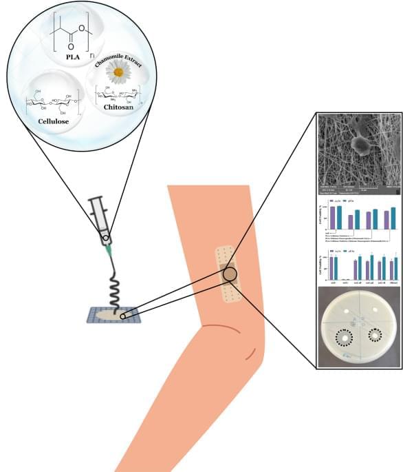
This study presents the development and characterization of a novel nanocomposite wound dressing material based on polylactic acid (PLA) nanofibers incorporating chitosan nanocapsules loaded with chamomile extract and cellulose nanoparticles.