Toggle light / dark theme

Circa 23 March 2020


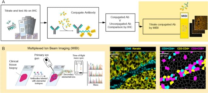
The ways in which a neoplastic cell arises and evades the immune system is the result of a departure from the systems biology that governs health. Understanding this biology requires methods that can resolve the heterogeneity of cell types, determine their states, whether they are activated (e.g., HLA-DR high) or suppressed (e.g., PD-1 high), and map their relationships or distances to one another. MIBI provides single cell resolution and sensitivity to phenotypically characterize the complex tissue environments including the TME. Executed similarly to IHC yet with the capability to profile 40+ markers simultaneously, MIBI is broadly applicable to a wide range of analyses performed in anatomic pathology including cell classification, spatial characterization, and assessment of marker expression. The MIBIscope produces data (multilayer TIFF files) that can be accessed by many analysis platforms currently available, such as those found in commercial software packages such as Fiji, Halo, and VisioPharm or freely available bioinformatic packages developed with open-source programming languages (e.g., R, Python).

All tumor types were stained, imaged, and analyzed using a single staining panel and standardized protocol. The workflow is flexible such that slides can be stained in batches and stored until imaged on the MIBIscope. Stained slides are typically stored under vacuum but protection from light is not necessary as the labels are stable metal isotopes rather than light-sensitive fluorophores. Once imaged it is possible to reimage the tissue as only a modest depth of the tissue is sputtered and analyzed during a single acquisition [16]. One limitation of the current project performed with an earlier version of the MIBIScope is the relatively small FOV size (500 μm by 500 μm) needed for images with 0.5 µm resolution. The current MIBIScope enables FOVs of 800 μm by 800 μm to be imaged in 70 min at fine resolution (650 nm). The resolution can be controlled at the instrument and acquisition at a slightly lower resolution than used in this study (1 μm) can be performed in 17 min. The 800 μm FOV captures 82% of a 1 mm TMA core. FOVs across cores of a TMA can be selected and then imaged in a single run. For whole sections it is possible to acquire adjacent images and stitch the images together using techniques commonly performed with other imaging technologies [22]. The need for tiling is particularly acute for imaging brain sections where multiple FOVs are collected to generate a larger image. Together with researchers at Stanford University, we are currently developing tiling methods to map large regions of brain tissue which will be described in a future publication. Because MIBI is still an early technology, the underlying methods for each stage of the processing pipeline are constantly evolving and improving, not just for accuracy but for generality. While the methods themselves are evolving, the pipeline tasks, at a high level, such as mass calibration, filtering, etc., are defined and have been automated through the MIBI/O software, and, as importantly, allows for appropriate user input when necessary. As more data becomes available, and the user base of MIBI grows, data processing should become more standardized.

The immediate utility of MIBI will be for understanding the biological mechanisms present in disease microenvironments. The results demonstrate the ability to detect a range of marker expression across many tumor types. The images can be segmented to define cell boundaries and then the expression of phenotypic markers used to classify cell instances into their cell class, such as proliferating tumor cells or nonproliferating tumor cells and various immune cells. Additional markers have been used on other sample sets to further define myeloid cell subsets, B cell subsets and stromal elements including vascular endothelial cells. This study also demonstrated the possibilities for calculating distances between different cell subsets including tumor and immune cells in addition to PD-1 and PD-L1 expressing immune cell subsets.

A genomic analysis of lung cancer in people with no history of smoking has found that a majority of these tumors arise from the accumulation of mutations caused by natural processes in the body. This study was conducted by an international team led by researchers at the National Cancer Institute (NCI), part of the National Institutes of Health (NIH), and describes for the first time three molecular subtypes of lung cancer in people who have never smoked.

These insights will help unlock the mystery of how lung cancer arises in people who have no history of smoking and may guide the development of more precise clinical treatments. The findings were published September 6 2021, in Nature Genetics.

“What we’re seeing is that there are different subtypes of lung cancer in never smokers that have distinct molecular characteristics and evolutionary processes,” said epidemiologist Maria Teresa Landi, M.D., Ph.D., of the Integrative Tumor Epidemiology Branch in NCI’s Division of Cancer Epidemiology and Genetics, who led the study, which was done in collaboration with researchers at the National Institute of Environmental Health Sciences, another part of NIH, and other institutions. “In the future we may be able to have different treatments based on these subtypes.”

To Sheng-Ying Pao, the power of reframing CRISPR lies in what is absolutely ordinary: paper. In CRISPaper, Pao revisited a cultural past in the ancient art of papermaking.

Over thousands of years, farmers painstakingly converted the wild rice plant into a staple crop. Today, researchers are using CRISPR to change genes to optimize grain yield. However, rice is more than food. In ancient China, it was used to make paper.

Pao took rice stalks from plants edited with CRISPR and ground the fibers into pulp. She then poured the pulp over a mesh screen. Every time she dipped the screen into water, the plant fibers would lift and resettle on top of the mesh, eventually making paper. Through the genome-edited rice plant, an ancient practice was juxtaposed with cutting-edge technology. Pao’s meditative ritual of papermaking is a counterbalance to the strangeness of the source material.
[Show ID: 37388]

https://news.berkeley.edu/story_jump/crispaper-understanding…rough-art/

Australians with COVID-19 who are at risk of hospitalisation will now have access to an additional antibody treatment, as the Therapeutic Goods Administration (TGA) announced today it has granted provisional approval for sotrovimab to be used in Australia.

Earlier this month, the Australian Government secured an initial allocation of over 7,700 doses of the novel monoclonal antibody treatment sotrovimab and a first shipment is already in the country and ready to be deployed through the National Medical Stockpile from next week.

The sotrovimab treatment requires a single dose to be administered through an intravenous (IV) infusion in a health care facility and has been shown to reduce hospitalisation or death by 79 per cent in adults with mild to moderate COVID-19, who are at risk of developing severe COVID-19.

The cumulative effect of reduced PTPN2 activity on both mechanisms was an elevated fluid loss. The researchers proved this defect could be reversed by treating cells lacking PTPN2 with recombinant -; or synthetic -; matriptase.


A team of researchers led by a biomedical scientist at the University of California, Riverside, has identified a novel mechanism by which loss-of-function mutations in the gene PTPN2, found in many patients with inflammatory bowel disease, or IBD, affect how intestinal epithelial cells maintain a barrier.

The intestinal epithelium, a single layer of cells, plays a critical role in human health by providing a barrier while also allowing nutrient and water absorption. Intestinal epithelial cells are needed for regulating immune function, communicating with the intestinal microbiota, and protecting the gut from pathogen infection -; all of which critically depend on an intact epithelial barrier.

Affecting roughly 3 million Americans, IBD is a set of chronic intestinal diseases in which the lining of the gut becomes inflamed and leaky. Increased gut leakiness has recently been confirmed to increase the risk of developing IBD.

More than 18 months into the COVID-19 pandemic, there are still very few drugs authorized by the FDA to treat the virus. Just one drug, remdesivir, has gained full FDA approval as a treatment for COVID-19.

Becker’s asked physicians from top health systems which medications they’re using most frequently to treat their COVID-19 patients, which they’re avoiding and which have been most effective.

Graphene, hexagonally arranged carbon atoms in a single layer with superior pliability and high conductivity, could advance flexible electronics according to a Penn State-led international research team. Huanyu “Larry” Cheng, Dorothy Quiggle Career Development Professor in Penn State’s Department of Engineering Science and Mechanics (ESM), heads the collaboration, which recently published two studies that could inform research and development of future motion detection, tactile sensing and health monitoring devices.

Investigating how laser processing affects graphene form and function

Several substances can be converted into carbon to create graphene through . Called laser-induced graphene (LIG), the resulting product can have specific properties determined by the original material. The team tested this process and published their results in SCIENCE CHINA Technological Sciences.

Two senior leaders in the US Food and Drug Administration’s vaccine review office are stepping down, even as the agency works toward high-profile decisions around Covid-19 vaccine approvals, authorizations for younger children and booster shots.

The retirements of Dr. Marion Gruber, director of the Office of Vaccines Research and Review at FDA’s Center for Biologics Evaluation and Research, and Dr. Philip Krause, deputy director of the office, were announced in an internal agency email sent on Tuesday and shared with CNN by the FDA.

In the email, CBER Director Dr. Peter Marks said Gruber will retire on October 31 and Krause is leaving in November. Marks thanked Gruber for her leadership throughout efforts to authorize and approve Covid-19 vaccines, and Krause for serving in a “key role in our interactions to address critical vaccine-related issues with our public health counterparts around the world.”

Why not eradicate disease for everyone?


Zolgensma – which treats spinal muscular atrophy, a rare genetic disease that damages nerve cells, leading to muscle decay – is currently the most expensive drug in the world. A one-time treatment of the life-saving drug for a young child costs US$2.1 million.

While Zolgensma’s exorbitant price is an outlier today, by the end of the decade there’ll be dozens of cell and gene therapies, costing hundreds of thousands to millions of dollars for a single dose. The Food and Drug Administration predicts that by2025it will be approving 10 to 20 cell and gene therapies every year.

I’m a biotechnology and policy expert focused on improving access to cell and gene therapies. While these forthcoming treatments have the potential to save many lives and ease much suffering, health care systems around the world aren’t equipped to handle them. Creative new payment systems will be necessary to ensure everyone has equal access to these therapies.

The study investigated whether electrical therapy, coupled with exercise, would show promise in treating tendon disease or ruptures. It showed that tendon cell function and repair can be controlled through electrical stimulation from an implantable device which is powered by body movement.


Researchers at CÚRAM, the SFI Research Centre for Medical Devices based at NUI Galway, have shown how the simple act of walking can power an implantable stimulator device to speed up treatment of musculoskeletal diseases.

The results of have been published in the prestigious journal Advanced Materials.

The research establishes the engineering foundations for a new range of stimulator devices that enable control of musculoskeletal tissue regeneration to treat tendon damage and disease and sports injuries, without the use of drugs or external stimulation.