“This led us to the following idea: what if instead of preventing the formation of droplets, we created conditions that would drive Tau and alpha synuclein inside the droplets toward their healthy path, discouraging them from taking the disease path?” said a co-corresponding author of the work.
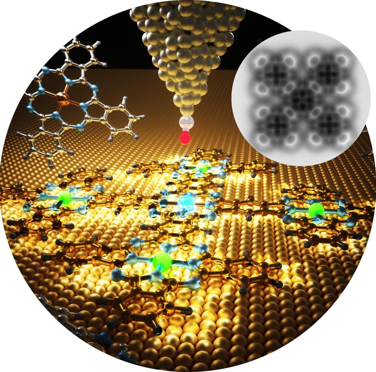
The team worked with biochemical and biophysical techniques, high-resolution microscopy and neuronal-based assays to investigate tubulin’s role in modulating and preventing the formation of toxic aggregates in droplets.
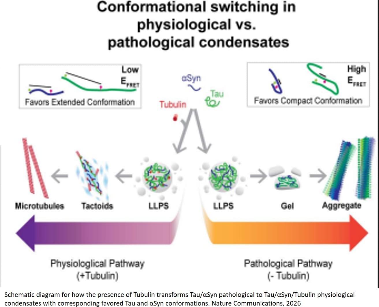
The researchers show that Tubulin modulates Tau:αSyn condensates by promoting microtubule interactions and inhibiting homotypic and heterotypic pathological oligomers. Tubulin partitioning into condensates promotes microtubule polymerization and prevents Tau and αSyn oligomerization.
In the absence of Tubulin, Tau-driven condensation accelerates formation of pathogenic Tau:αSyn heterodimers and amyloid fibrils. The authors also identify distinct Tau and αSyn structural states in pathological Tubulin-absent versus physiological Tubulin-rich condensates.
“When tubulin levels are low, as it has been found in Alzheimer’s disease, microtubules are less abundant and Tau and alpha synuclein can form toxic aggregates,” the author said. “But when tubulin is present, Tau and alpha‑synuclein shift away from harmful aggregates and instead promote the assembly of healthy microtubules,” the author said. “Tubulin redirects the activity of these proteins by giving them something productive to do.” ScienceMission sciencenewshighlights.
Researchers have discovered a potential new strategy to fight back against Alzheimer’s and Parkinson’s diseases, conditions that are linked to the toxic accumulation of Tau and alpha synuclein protein clumps in the brain. The team reports in Nature Communications that tubulin, the building block of microtubules, the cell’s internal ‘railway tracks,’ can stop Tau and alpha synuclein from forming toxic clumps and instead steer them into their normal, healthy roles.