Toggle light / dark theme

Frontiers: Biological membranes are complex, heterogeneous, and dynamic systems that play roles in the compartmentalization and protection of cells from the environment

It is still a challenge to elucidate kinetics and real-time transport routes for molecules through biological membranes in live cells. Currently, by developing and employing super-resolution microscopy; increasing evidence indicates channels and transporter nano-organization and dynamics within membranes play an important role in these regulatory mechanisms. Here we review recent advances and discuss the major advantages and disadvantages of using super-resolution microscopy to investigate protein organization and transport within plasma membranes.

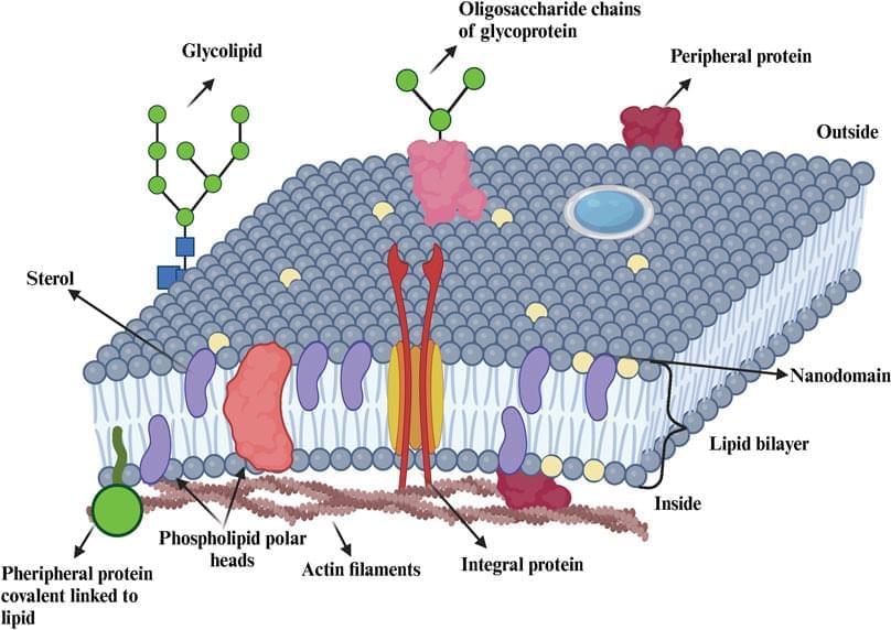
The mammalian plasma membrane (PM) is a complex assembly of lipids and proteins that separates the cell’s interior from the outside environment (Ingolfsson et al., 2014). The multiple collective processes that take place within membranes have a strong impact not only on the cellular behavior but also on its biochemistry. Understanding these processes poses a challenge due to the often complex and multiple interactions among membrane components (Stone et al., 2017). Moreover, the PM surface accommodates different types of lipid and protein clusters (Saka et al., 2014; Owen et al., 2012; Sezgin, 2017), even though the functional role of the clustering on the membrane surface has not yet been fully understood.

First-principles approaches and concepts to simulate electrochemical interfaces

State-of-the-art approaches for modelling electrified solid–electrolyte interfaces are critically discussed, highlighting key challenges in incorporating thermodynamic open-boundary conditions, large electrostatic potentials and their dynamic fluctuations into realistic ab initio simulations.

GOOD LUCK, HAVE FUN, DON’T DIE — Welcome To The Perfect Prison

Gore Verbinski’s Good Luck, Have Fun, Dont Die hits like a nasty mirror held up at the worst possible angle. On paper, the setup sounds almost playful: a “Man From the Future” drops into a diner in Los Angeles and has to recruit the exact combination of disgruntled strangers for a one-night mission to stop a rogue AI. But the horror isn’t metal skeletons and laser fire. It’s the idea that the end of humanity doesn’t arrive with an explosion. It arrives with an upgrade. A perfectly tuned stream of algorithmic entertainment that doesn’t merely distract people—it replaces them. A manufactured paradise so frictionless, so gratifying, so chemically rewarding, that the messy, strenuous, inconvenient act of being human starts to feel obsolete.

#goodluckhavefundontdie #samrockwell #ai #algorithm.

Check out my playlists on film here — Film Explored — • Film Explored.

Check out my playlist on Alien here — • New to Aliens? Start Here.

Check out my playlist on Predator here — • New to Predator? Start Here.

Stick chemistry: High-throughput method for finding new molecular glues

Molecular glues are enjoying the spotlight, but discovering new ones is often a matter of luck. A new method, developed by Scripps Research chemist Michael Erb and colleagues, aims to discover new glues more intentionally with a “target-based” approach.


Click chemistry method opens up avenues for intentional glue discovery by .

Chemistry-powered ‘breathing’ membrane opens and closes tiny pores on its own

Ion channels are narrow passageways that play a pivotal role in many biological processes. To model how ions move through these tight spaces, pores need to be fabricated at very small length scales. The narrowest regions of ion channels can be just a few angstroms wide, about the size of individual atoms, making reproducible and precise fabrication a major challenge in modern nanotechnology.

In a study published in Nature Communications, researchers at The University of Osaka have addressed this challenge by using a miniature electrochemical reactor to create ultra-small pores approaching subnanometer dimensions.

In biological cells, ions flow in and out through channels in cell membranes. This ion flow is the basis for generating electrical signals, such as nerve impulses that trigger muscle contraction. The channels themselves are made of proteins and can have angstrom-wide narrow regions. Conformational changes of these proteins in response to external stimuli open and close the channels.

Role of Dopamine in Pain

Dopamine is a member of a class of molecules called the catecholamines, which serve as neurotransmitters and hormones. In the brain, dopamine serves as a neurotransmitter and is released from nerve cells to send signals to other nerves. Outside of the nervous system, it acts as a local chemical messenger in several parts of the body.

Image Copyright: Meletios, Image ID: 71,648,629 via shutterstock.com

A number of important neurodegenerative diseases are associated with abnormal function of the dopamine system and some of the main medications used to treat those illnesses work by changing the effects of dopamine. The condition Parkinson’s disease is caused by a loss of dopamine secreting cells in a brain area called the substantia nigra.

Supercomputer simulations reveal rotation drives chemical mixing in red giant stars

Advances in supercomputing have made solving a long‐standing astronomical conundrum possible: How can we explain the changes in the chemical composition at the surface of red giant stars as they evolve?

For decades, researchers have been unsure exactly how the changing chemical composition at the center of a red giant star, caused by nuclear burning, connects to changes in composition at the surface. A stable layer acts as a barrier between the star’s interior and the outer connective envelope, and how elements cross that layer remained a mystery.

In a Nature Astronomy paper, researchers at the University of Victoria’s (UVic) Astronomy Research Center (ARC) and the University of Minnesota solved the problem.

Quantum trembling: Why there are no truly flat molecules

Traditional chemistry textbooks present a tidy picture: Atoms in molecules occupy fixed positions, connected by rigid rods. A molecule such as formic acid (methanoic acid, HCOOH) is imagined as two-dimensional—flat as a sheet of paper. But quantum physics tells a different story. In reality, nature resists rigidity and forces even the simplest structures into the third dimension.

Researchers led by Professor Reinhard Dörner of the Institute for Nuclear Physics at Goethe University have now determined the precise spatial structure of the “flat” formic acid molecule using an X-ray beam from the PETRA III synchrotron radiation source at the DESY accelerator center in Hamburg. They collaborated with colleagues from the universities of Kassel, Marburg and Nevada, the Fritz Haber Institute, and the Max Planck Institute for Nuclear Physics. The study is published in Physical Review Letters.

To accomplish this, they made use of two effects that occur when X-ray radiation strikes a molecule. First, the radiation ejects several electrons from the molecule (photoelectric effect and Auger effect). As a result, the atoms become so highly charged that the molecule bursts apart in an explosion (Coulomb explosion). The scientists succeeded in measuring these processes sequentially, even though they take place within femtoseconds—millionths of a billionth of a second.

/* */