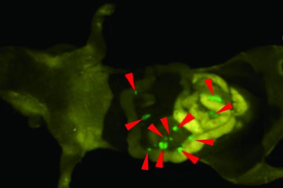
Toggle light / dark theme

Patients with certain types of early stage cancer, particularly those affecting the gastrointestinal system, may be able to avoid surgery and be successful

Imagine a world in which free-floating electric vehicles charge wirelessly as they glide down highways, laptops are hundreds of times more powerful, and clean energy flows in limitless supply.

Such a future, experts say, hinges on the development of new superconductors, or materials capable of transmitting electricity with near-perfect efficiency. The problem? All known superconductors—from pure elements like lead, tin, and aluminum to exotic compounds like niobium–titanium—must be subjected to or pressure to function, making them impractical for widespread use. More problematic still, scientists don’t fully understand how these materials work, making it difficult to engineer better versions.

Superconductors have already made their way into MRI machines, particle accelerators, and electromagnetic levitating trains, but they are extraordinarily expensive and finicky. The real game changer, experts say, will be figuring out how to custom-design superconductors that are cheaper and more versatile.

Individuals with retinal degenerative diseases struggle to restore vision due to the inability to regenerate retinal cells. Unlike cold-blooded vertebrates, mammals lack Müller glia (MG)-mediated retinal regeneration, indicating the limited regenerative capacity of mammalian MG. Here, we identify prospero-related homeobox 1 (Prox1) as a key factor restricting this process. Prox1 accumulates in MG of degenerating human and mouse retinas but not in regenerating zebrafish. In mice, Prox1 in MG originates from neighboring retinal neurons via intercellular transfer. Blocking this transfer enables MG reprogramming into retinal progenitor cells in injured mouse retinas. Moreover, adeno-associated viral delivery of an anti-Prox1 antibody, which sequesters extracellular Prox1, promotes retinal neuron regeneration and delays vision loss in a retinitis pigmentosa model. These findings establish Prox1 as a barrier to MG-mediated regeneration and highlight anti-Prox1 therapy as a promising strategy for restoring retinal regeneration in mammals.


Recovery for mammalian retinal degeneration is limited by a lack of Müller glia (MG)-mediated regeneration. Here authors show blocking Prox1 accumulation and intercellular transfer from retinal neurons enables MG reprogramming of retinal progenitor cells, promotes retinal neuron regeneration, and delays vision loss.

Urban rats spread a deadly bacteria as they migrate within cities that can be the source of a potentially life-threatening disease in humans, according to a six-year study by Tufts University researchers and their collaborators that also discovered a novel technique for testing rat kidneys.

Leptospirosis is a disease caused by a type of bacteria often found in rats. It’s spread through their urine into soil, water, or elsewhere in the environment, where it becomes a source of infection and contamination for humans, dogs, and other species. While it’s prevalent worldwide, it’s more common in tropical regions, though a changing climate means it could become more common in colder regions as they warm.

In Boston, leptospirosis persists in local rat populations, and different strains of the bacteria move around the city as groups of rats migrate, according to a new study by Marieke Rosenbaum, M.P.H., D.V.M., assistant professor in the Department of Infectious Disease and Global Health at Cummings School of Veterinary Medicine at Tufts University, along with co-authors at Northern Arizona University (NAU), the United States Department of Agriculture (USDA), and the Centers for Disease Control and Prevention (CDC). In addition, their of a 2018 human leptospirosis case in Boston strongly suggests a link to rats as the source.

A McGill University-led research collaboration has achieved a breakthrough in understanding how cancer spreads. A clinical study of ovarian and colorectal cancer patients found cancer cells move in the bloodstream in clusters more commonly than was previously thought. The discovery could help doctors more quickly identify which cancer patients are at high risk of having their cancer spread to other organs, knowledge that could guide treatment decisions. The findings also potentially open new avenues for treatment.

The study, published in Communications Medicine, was conducted with researchers and clinicians Anne-Marie Mes-Masson and Dr. Diane Provencher at the Centre hospitalier de l’Université de Montréal, Dr. Peter Metrakos at the Research Institute of the McGill University Health Centre and Luke McCaffrey at the McGill-affiliated Rosalind and Morris Goodman Cancer Institute.

Cancer is responsible for about 1 in 4 deaths in Canada. In most cases, it is not the original tumor that proves fatal, but the cancer spreading to other organs, a process called metastasis. This occurs when circulating tumor cells (CTCs) break away from tumors, enter the bloodstream, and seed new tumors elsewhere in the body. On rare occasions, CTCs break away as a group of cells sticking to one another and forming a cluster.

The strong links between changes in astrocyte structure and function in the context of neurodevelopment and disease have been supported by studies examining astrocyte cytoskeletal markers such as glial fibrillary acidic protein (GFAP) in disease models and postmortem human brain tissue, where increases or decreases in its expression in various brain nuclei are often linked with neurocognitive and psychiatric disorders. Hence, changes in GFAP expression are often the first-line test for astrocyte involvement in disease and support a role for astrocyte dysfunction in major depression, schizophrenia, alcohol and substance use disorders, anorexia nervosa, and bipolar disorder (719), where changes in astrocyte structure, density, complexity, and/or blood vessel association are linked with disrupted astrocyte function. Although reactive astrogliosis remains the single most studied astrocytic response involving morphological adaptations and changes in GFAP expression (20, 21), in recent years, astrocyte morphological plasticity has been shown to be more nuanced. GFAP expression is dynamic across the circadian cycle (2224) and increases with physical exercise and environmental enrichment (25, 26). Moreover, in aging, astrocytes increase or decrease their GFAP expression in different brain regions (27, 28), suggesting heterogeneity in astrocyte form and function.

We previously found a notable relationship between astrocyte structure and vulnerability to substance use disorders, with astrocytes in the nucleus accumbens (NAc) altering their association with different neural subcircuits to drive or suppress drug-seeking behavior depending on heroin availability (2931). The NAc is critical for regulating behavioral outputs in response to rewards, including substances of abuse and natural reinforcers, such as food or sucrose. The NAc is composed of core and shell subregions that are themselves heterogeneous structures with regard to synaptic input and output connectivity and function (3236). Heterogeneity has been observed in astrocyte morphology within the NAc core (3, 30, 37), but studies have not yet examined how astrocyte structure and function differ across NAc subregions at baseline or in response to operant conditioning with natural or pathological reinforcers.

To address this gap, we developed an automated pipeline for single-cell morphological analysis of astrocytes that integrates state-of-the-art deep learning models for astrocyte detection and segmentation, together with highly sensitive geometrical tools for precise quantitation of single-cell morphological characteristics. We introduce the rigorous notion of morphological distance (MD) to measure alterations in astrocyte morphology and compare astrocyte subpopulations according to their structural characteristics. By applying this pipeline in combination with supervised machine learning, we found that single-astrocyte morphological characteristics were predictive not only of anatomical location within the NAc at baseline but also of the availability of heroin or sucrose at the moment of image capture. This geometrically sensitive approach yields substantially more detailed information about astrocyte structure than previously applied manual or semiautomated approaches and serves as a rigorous quantitative assay for identifying brain nuclei where astrocytes undergo plasticity in the context of disease. We found that astrocyte structural plasticity across the NAc was disrupted in animals that had been exposed to heroin but not sucrose, consistent with a largely protective role for NAc astrocytes in maintaining synaptic homeostasis and behavioral flexibility. We also found that astrocyte structural plasticity in the dorsomedial portion of the NAc shell was uniquely engaged during the initiation of opioid but not sucrose seeking, suggesting the involvement of this structure in drug relapse.

The researchers discovered that AP2A1 seemed to be responsible for switching cells between their “young” and “old” states—senescent cells were rejuvenated by the suppression of the protein, and younger cells aged by its overexpression.

The scientists also found that the AP2A1 was frequently in close proximity to another protein: integrin β1, which aids cells in binding to the collagen scaffold that envelops them. Both proteins, the researchers described, travel along stress fibers within cells.

Chinese researchers have developed a wireless, paper-thin patch that attaches to an organ to create a highway for drug delivery.

To solve an important problem in drug delivery, a research team that includes Beihang University and Peking University developed an electronic patch that acts like a band-aid for organs.

Traditional drug delivery systems send a vague package through the body that requires higher doses than necessary and might harm organs in the process of trying to find their destination. Large-molecule drugs, or biopharmaceuticals based on proteins, face an even greater challenge as the cell membrane often blocks these drugs, according to CGTN.