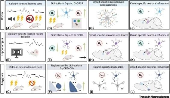
Toggle light / dark theme

‘Mesoscale’ swimmers could pave way for drug delivery robots inside the body

In physics, the mesoscale lies between the microscopic and the macroscopic. It is not just the domain of tiny living creatures like small larvae, shrimp, and jellyfish, but also where physics equations become extreme. While the macroscopic realm is governed by inertia and the microscopic by viscosity, the mesoscale is both and neither, requiring a new set of physics to describe it.

Now, physicists at Aalto University’s Department of Applied Physics have discovered how organisms swim in the mesoscale mix of viscosity and inertia. The study was recently published in the journal Communications Physics.

Led by Assistant Professor Matilda Backholm, the multidisciplinary team found the key to efficient swimming in this realm is not just moving faster or growing bigger, but a phenomenon of non-reciprocal motion known as time reversal symmetry breaking. The results help fill a knowledge gap in fundamental physics and could pave the way for applications such as mesorobotics; tiny robots injected inside a patient’s body for drug delivery or carrying out medical procedures.

Robotic microfluidic platform brings AI to lipid nanoparticle design

AI has designed candidate drugs for antibiotic-resistant infections and genetic diseases. But efforts to incorporate AI into the design of lipid nanoparticles (LNPs), the revolutionary delivery vehicles behind mRNA therapies like the COVID-19 vaccines, have been much more limited.

Designing LNPs is especially challenging: Each formulation combines multiple lipid components whose ratios influence how the particle delivers genetic instructions inside cells. Scientists still lack a clear map connecting those chemical inputs to biological outcomes.

The reason? There simply isn’t enough data.

Mapping 3D-super-enhancers with machine learning to pinpoint regulators of cell identity

Scientists usually study the molecular machinery that controls gene expression from the perspective of a linear, two-dimensional genome—even though DNA and its bound proteins function in three dimensions (3D). To better understand how key components of this machinery, such as super-enhancers, regulate genes in this 3D reality, scientists at St. Jude Children’s Research Hospital have developed a new algorithm called BOUQUET.

Using machine learning, BOUQUET reveals that sets of genes and their regulatory elements can interact within protein condensates, high-density membraneless droplets, in cells’ nuclei. The findings, which provide new insight into how cells regulate the genes that control their specialized identities, were published today in Nucleic Acids Research.

Cells express certain sets of genes to carry out specific functions; for example, a blood cell and a brain cell express different context-specific genes. There are 3 billion base pairs of human DNA, and the genes involved in cell identity are scattered throughout. Even more challenging, enhancers, DNA elements that activate gene expression, can be thousands of DNA bases away from their target genes.

Malicious npm Package Posing as OpenClaw Installer Deploys RAT, Steals macOS Credentials

Cybersecurity researchers have discovered a malicious npm package that masquerades as an OpenClaw installer to deploy a remote access trojan (RAT) and steal sensitive data from compromised hosts.

The package, named “@openclaw-ai/openclawai,” was uploaded to the registry by a user named “openclaw-ai” on March 3, 2026. It has been downloaded 178 times to date. The library is still available for download as of writing.

JFrog, which discovered the package, said it’s designed to steal system credentials, browser data, crypto wallets, SSH keys, Apple Keychain databases, and iMessage history, as well as install a persistent RAT with remote access capabilities, SOCKS5 proxy, and live browser session cloning.

Chemistry in Pictures: Glowing with pride

“It looks like a tiny solar system. But instead of planets, it’s a snapshot of my research journey in the lab,” says Sadiya Tanga, a chemistry graduate student at Ashoka University. Tanga’s work has focused on a type of drug molecule called proteolysis-targeting chimeras, or PROTACs for short. PROTACs have two active ends, one that grabs a target protein and another that grabs a molecular flag that tells the cell to break down the whole assembly as waste. “Each glowing flask and sphere holds a different compound I worked hard to design and synthesize,” Tanga says. “The colors you see are from parts of the molecules that shine under UV light.”

Neuroimaging Biomarkers of Disease Progression and Cognitive Change in Patients With Retinal Vasculopathy With Cerebral Leukoencephalopathy

The official journal of the Guarantors of Brain. Provides researchers and clinicians with original contributions in neurology by publishing a wide range of original studies in neurological science, in addition to practical clinical articles.

Scientists discover hidden brain cells that may stop Alzheimer’s tau buildup

Scientists have uncovered a surprising new role for little-known brain cells called tanycytes that may influence the development of Alzheimer’s disease. These specialized cells appear to help remove toxic tau protein from the brain by transporting it from the cerebrospinal fluid into the bloodstream. When tanycytes become damaged or dysfunctional, tau can accumulate in the brain, a hallmark of Alzheimer’s.

Circulating MALAT1 in Preeclampsia and Association With Cardiometabolic Risk

RESEARCH ARTICLE: Circulating MALAT1 in Preeclampsia and Association With Cardiometabolic Risk @tovelekva


BACKGROUND: Preeclampsia is a hypertensive disorder affecting 2% to 8% of pregnancies. Women with a history of preeclampsia have an increased risk of cardiovascular disease. The long noncoding RNA MALAT1 is shown to regulate inflammatory responses linked to cardiovascular disease. MALAT1 is decreased in preeclampsia placentas and may have a cis-regulatory function on neighboring RNAs. METHODS: Expression of MALAT1, NEAT1, mascRNA, SCYL1, and FRMD8 was assessed in peripheral blood mononuclear cells, and MALAT1 in plasma and extracellular vesicles, at 22 to 24 and 36 to 38 weeks of gestation in healthy (n=214) and preeclampsia (n=37) women from the STORK cohort study (STORe barn og Komplikasjoner, translated as Large Babies and Complications) and at 5-year follow-up in women with and without history of preeclampsia (n=29; n=271).

Recent pandemic viruses jumped to humans without prior adaptation, study finds

A new University of California San Diego study published in Cell challenges a long-standing assumption about how animal viruses become capable of sparking human epidemics and pandemics. Using a phylogenetic, genome-wide analysis across multiple viral families, researchers report that most zoonotic viruses—infectious pathogens that spread from animals to humans, including the cause of COVID-19—do not show evidence of special evolutionary adaptation before spilling over into humans.

“This work has direct relevance to the ongoing controversy around COVID-19 origins,” said Joel Wertheim, Ph.D., senior author and professor of medicine in the Division of Infectious Diseases and Global Public Health at UC San Diego School of Medicine.

“From an evolutionary perspective, we find no evidence that SARS-CoV-2 was shaped by selection in a laboratory or prolonged evolution in an intermediate host prior to its emergence. That absence of evidence is exactly what we would expect from a natural zoonotic event—and it represents another nail in the coffin for theories invoking laboratory manipulation.”

/* */