The first human clinical trial of a universal Sarbeco coronavirus vaccine, developed by the University of Cambridge and spin-out DIOSynVax (DVX) Ltd, has shown that the vaccine is safe and has no significant side effects.
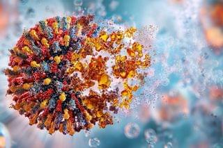
The trial, involving 39 healthy volunteers, tested a vaccine designed to provide protection against multiple Sarbeco coronaviruses—the large group of viruses that occur in nature including SARS-CoV-2, which caused the COVID pandemic.
The vaccine triggered immune responses in the volunteers not only to SARS-CoV-2 and SARS, but to related bat viruses that could potentially jump from animals to humans and cause future pandemics.
