Researchers have identified a vitamin B12–based compound that appears capable of crossing the blood–brain barrier and selectively accumulating in glioblastoma tissue.

From that insight, Dirac built an entirely new formulation of the theory using what he called “q-numbers” (quantum numbers)—abstract quantities that don’t commute. He independently rediscovered aspects of Hilbert’s operator theory, though he preferred his own algebraic route because he found mathematicians’ obsession with convergence and existence theorems unappealing.
Paul Adrien Maurice Dirac (, dih-RAK ; [ 3 ] 8 August 1902 – 20 October 1984) was a British theoretical physicist who is considered to be one of the founders of quantum mechanics. [ 4 ] [ 5 ] Dirac laid the foundations for both quantum electrodynamics and quantum field theory, coining the former term. [ 6 ] [ 7 ] [ 8 ] [ 9 ] He was Lucasian Professor of Mathematics at the University of Cambridge from 1932 to 1969, and a professor of physics at Florida State University from 1970 to 1984. Dirac shared the 1933 Nobel Prize in Physics with Erwin Schrödinger “for the discovery of new productive forms of atomic theory.” [ 10 ]
Dirac graduated from the University of Bristol with a Bachelor of Science in Electrical Engineering in 1921, and a Bachelor of Arts in Mathematics in 1923. [ 11 ] Dirac then graduated from St John’s College, Cambridge, with a Doctor of Philosophy in Physics in 1926, writing the first ever thesis on quantum mechanics. [ 12 ]
He formulated the Dirac equation, one of the most important results in physics, in 1928. [ 7 ] It connected special relativity and quantum mechanics and predicted the existence of antimatter. [ 13 ] He wrote a famous paper in 1931, [ 14 ] which further predicted the existence of antimatter. [ 15 ] [ 16 ] [ 13 ] Dirac also contributed greatly to the reconciliation of general relativity with quantum mechanics. He contributed to Fermi–Dirac statistics, which describes the behaviour of fermions, particles with half-integer spin. His 1930 monograph, The Principles of Quantum Mechanics, is one of the most influential texts on the subject. [ 17 ] He and Schrödinger tied for eighth in a Physics World poll of the greatest physicists of all time. [ 18 ] .
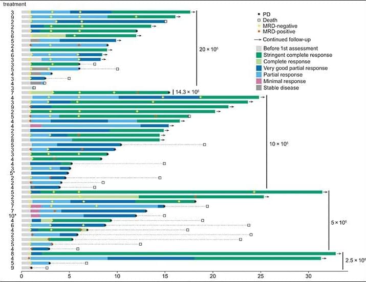
Prolonged manufacturing times for autologous CAR T cell therapies can be incompatible with rapidly progressive disease (PD), resulting in increased need for bridging therapy to achieve disease stabilization. Bridging therapy was required for most patients receiving cilta-cel and ide-cel in clinical trials (75 and 87%, respectively) (7, 9, 11, 12). Although use of bridging therapy may not affect ORR, CRR, or PFS, it is associated with worse overall survival (15). Similarly, as wait times for CAR T cell product increase, so does risk of mortality as effectiveness of the therapy decreases (16, 17), highlighting the need for improved CAR T cell products with faster and more reliable manufacturing.
Another issue associated with traditionally manufactured CAR T cell products is T cell exhaustion due to extended periods of in vitro stimulation and expansion during manufacturing (18). Higher levels of exhausted T cells were also observed in the leukapheresis material and final products from patients who later experienced PD (18). T cell exhaustion can result in poor persistence of CAR T cells in the body, thereby impeding function as the proliferation and survival of transferred T cells strongly correlate with their antitumor activity (19–22). Specific T cell populations have varying abilities to expand and persist in vivo. Memory (CD8+CD45RO−CD27+) and naive T cell (TN cell) subsets are associated with improved clinical response, given their ability to proliferate and persist after infusion, whereas effector T cell subsets comparatively exhibit lower self-renewal and survival capabilities (19, 23, 24). Although these patient-specific parameters are initially established in leukapheresis material, preservation of such cell populations in the final product via manufacturing techniques may improve the antitumor activity of a patient’s CAR T cell therapy (18, 19, 23, 24).
Durcabtagene autoleucel (PHE885) is an autologous, BCMA-targeting CAR T cell therapy carrying a CAR construct with a fully human anti-BCMA single-chain fragment variable (scFv) fused to 4-1BB/CD3ζ signaling domains manufactured on a next-generation platform. Prior work has shown that this platform can successfully manufacture product in fewer than 2 days by eliminating the need for ex vivo expansion, thereby preserving overall T cell stemness (the ability of T cells to self-renew and mature), which results in a final product with greater proliferative potential and fewer exhausted T cells (18). Here, we present the findings of part A of the phase 1 study (NCT04318327) of durcabtagene autoleucel in r/r MM, along with correlative analyses of the product before and after infusion.

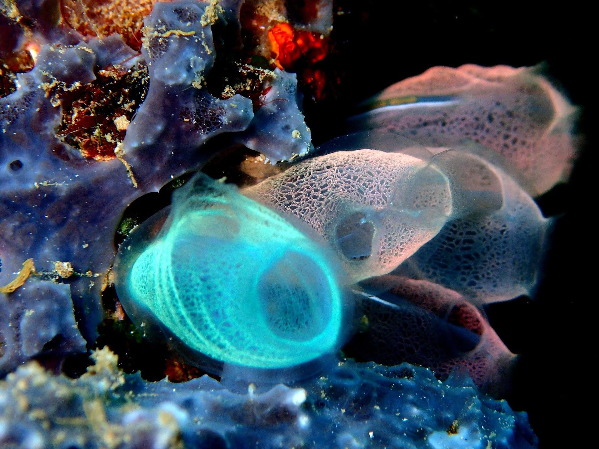
A tiny sea creature might hold the secret to reversing the aging process. When treated with a brief series of electrical pulses, sea squirts experience dramatic and long-lasting health improvements that can significantly extend their lifespans, according to a new study by researchers at Stanford and other institutions.
The findings, published in PNAS, open new possibilities for protecting marine species from warming waters, learning what causes stem cells in our own bodies to degrade, and potentially finding new ways to use these cells to treat medical conditions.
“This treatment recharges stem cells,” said study co-senior author Ayelet Voskoboynik, an assistant professor of biology in the Stanford School of Humanities and Sciences. “Understanding this mechanism is the key to unlocking how we might one day slow stem cell aging and trigger rejuvenation pathways.”
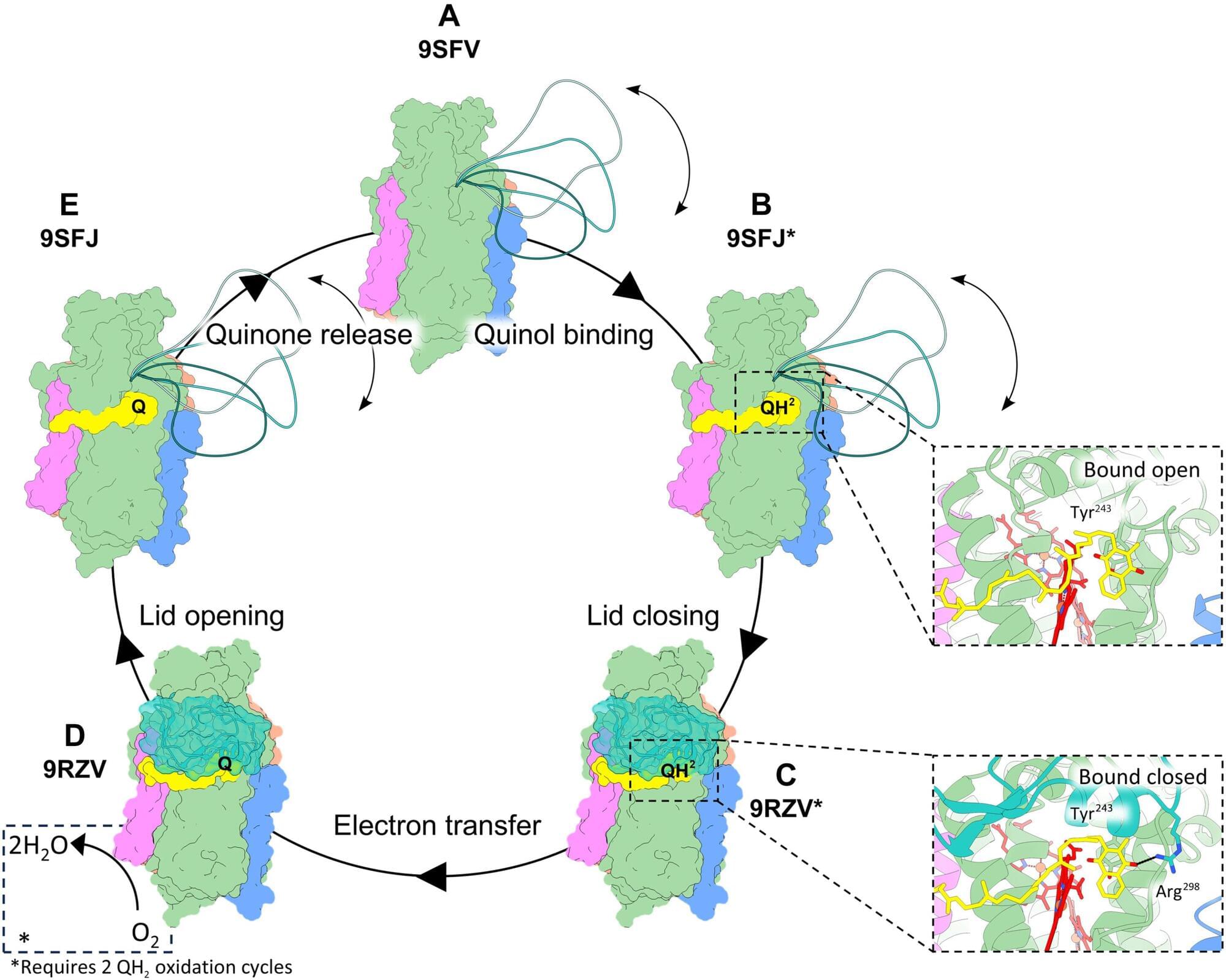
Researchers in Leiden have, for the first time, observed how a specialized enzyme helps bacteria stay alive when oxygen levels are low, and how that process can be blocked. The study, published in Science Advances, opens up new possibilities for targeted antibiotics.
It really exists: a secret trick that allows bacteria to survive with very little oxygen. This also applies to bacteria that can make us sick. “Like us, these bacteria need oxygen to survive,” says Ph.D. candidate Tijn van der Velden. “But unlike humans, they have a special enzyme called cytochrome bd that allows them to keep producing energy even when oxygen levels are very low.” Because the enzyme is so important for bacterial survival, it is a promising target for new antibiotics, including potential treatments for tuberculosis.

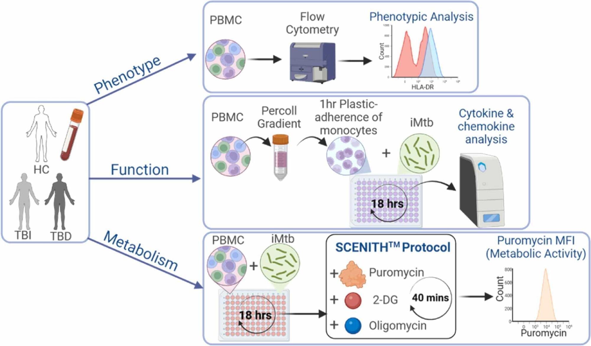
Researchers at Trinity College Dublin have identified key differences in how immune cells generate and use energy, a process known as cellular metabolism, in people with latent versus active tuberculosis (TB). The findings offer new insights into why some individuals control infection while others develop disease.
The study, published in the Journal of Infection, focused on circulating monocytes, key immune cells involved in the defense against TB infection. The researchers found that cells from people with latent TB remain metabolically flexible, allowing them to mount strong antibacterial responses, whereas cells from people with active TB disease show impaired metabolism and weaker responses to infection.
TB remains the world’s leading infectious killer, with 10.8 million cases and 1.25 million deaths recorded globally in 2023. While many people infected with Mycobacterium tuberculosis never become ill, researchers still do not fully understand why some individuals progress to active disease while others successfully control the infection. The findings could help pave the way for improved TB monitoring tools and future therapies or vaccines that target how immune cells generate energy.