Toggle light / dark theme

You could say that Illumina is to DNA sequencing is what Google is to Internet search, but that would be underselling the San Diego-based biotech company. Illumina’s machines, the best and cheapest on the market, generate 90 percent of all DNA sequence data today. Illumina is, as they say, crushing it.

But as lucrative as that 90 percent slice is for Illumina now, the whole pie is likely to get even bigger in the future. Less than 0.01 percent of the world’s population has been sequenced so far. So recently, Illumina has made bold moves positioning itself for the future: The company is consolidating its core hardware business—this week, it sued an upstart competitor, Oxford Nanopore Technologies, for patent infringement—while moving into the genetic testing business with new ventures like the liquid cancer biopsy spinoff, Grail.

The company is a looking toward a future in which a lot more people gets genetic tests—and a lot more often. “Grail’s business will be very different than Illumina’s core business,” Eric Endicott, Illumina’s director of global public relations, said in an email. “We are at a tipping point in genomics, where a broad community of scientists and researchers continue to translate the potential of the genome from science to discoveries and applications.”

Read more

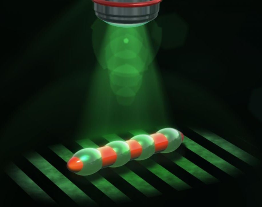
Ciliates can do amazing things: Being so tiny, the water in which they live is like thick honey to these microorganisms. In spite of this, however, they are able to self-propel through water by the synchronized movement of thousands of extremely thin filaments on their outer skin, called cilia. Researchers from the Max Planck Institute for Intelligent Systems in Stuttgart are now moving robots that are barely perceptible to the human eye in a similar manner through liquids. For these microswimmers, the scientists are neither employing complex driving elements nor external forces such as magnetic fields. The team of scientists headed by Peer Fischer have built a ciliate-inspired model using a material that combines the properties of liquid crystals and elastic rubbers, rendering the body capable of self-propelling upon exposure to green light. Mini submarines navigating the human body and detecting and curing diseases may still be the stuff of science fiction, but applications for the new development in Stuttgart could see the light-powered materials take the form of tiny medical assistants at the end of an endoscope.

Their tiny size makes life extremely difficult for swimming microorganisms. As their movement has virtually no momentum, the friction between the water and their outer skin slows them down considerably — much like trying to swim through thick honey. The viscosity of the medium also prevents the formation of turbulences, something that could transfer the force to the water and thereby drive the swimmer. For this reason, the filaments beat in a coordinated wave-like movement that runs along the entire body of the single-celled organism, similar to the legs of a centipede. These waves move the liquid along with them so that the ciliate — measuring roughly 100 micrometres, i.e. a tenth of a millimetre, as thick as a human hair — moves through the liquid.

“Our aim was to imitate this type of movement with a microrobot,” says Stefano Palagi, first author of the study at the Max Planck Institute for Intelligent Systems in Stuttgart, which also included collaborating scientists from the Universities of Cambridge, Stuttgart and Florence. Fischer, who is also a Professor for Physical Chemistry at the University of Stuttgart, states that it would be virtually impossible to build a mechanical machine at the length scale of the ciliate that also replicates its movement, as it would need to have hundreds of individual actuators, not to mention their control and energy supply.

Read more

African trypanosomiasis is called ‘sleeping sickness’ because when the infection is untreated, trypanosome parasites will invade the brain and cause disruption of sleeping patterns and irreversible neurological damage. A study published on February 25th in PLOS Pathogens reports that in a mouse model of trypanosome disease, nitric oxide (NO) plays an unexpected role in preserving the integrity of the blood brain barrier (BBB), thereby reducing parasite invasion into the brain, and likely limiting neurological damage.

NO is generally thought to be a pro-inflammatory signal, promoting a strong immune response against pathogens. The resulting inflammation is a mixed blessing: on one hand, it helps to control potentially dangerous pathogens, but on the other, it can cause “collateral damage” to the inflamed tissue.

Martin Rottenberg and colleagues, from the Karolinska Institute in Stockholm, Sweden, are interested in how trypanosome cause disease and in the host immune defense against them. In this study, they examine the role of NO in a of trypanosomiasis, with a focus on how the parasites manage to get through the so-called (BBB), the border surrounding the mammalian that is normally impenetrable to foreign intruders as well as most host cells.

Read more

Just Amazing


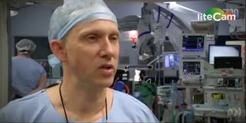
Ralph Mobbs, a neurosurgeon at the Prince of Wales Hospital in Sydney, made medical history in late 2015 when he successfully replaced two vertebrae with custom made prosthesis. The patient, in his 60s, suffered from Chordoma, a particularly nasty form of cancer that had formed on his top two vertebrae and threatened to cinch off his spinal cord as it grew. That would have left him a quadriplegic. Complicating matters, those top two vertebrae are what allow you to turn and tilt your head, so it’s not like doctors can easily fashion a replacement out of bone grafted from another part of the patient’s body. They have to fit perfectly and that’s where the 3D printers come in.

Mobbs worked with Anatomics, an Australian medical device manufacturer, to craft perfect replicas of the patient’s top two vertebrae out of titanium. This is the first time that these two particular neck bones have been printed and installed. “To be able to get the printed implant that you know will fit perfectly because you’ve already done the operation on a model … It was just a pure delight,” Mobbs told Mashable Australia. “It was as if someone had switched on a light and said ‘crikey, if this isn’t the future, well then I don’t know what is’.”

The surgery itself was no small feat. The 15-hour procedure is fraught with peril as the medical team operates within inches of the top of the spinal cord as well as the brainstem and numerous major arteries. “The surgery that we’re doing today is a particularly complicated and long and difficult surgery. It involves exposure at the top of the neck where the neck and the head meets,” Mobbs told ABC 7.30. “It’s essentially disattaching the patient’s head from his neck and taking the tumour out and reattaching his head back onto his neck.” Thankfully, the surgery was a success. Mobbs was able to remove the tumor and implant the prosthetic.

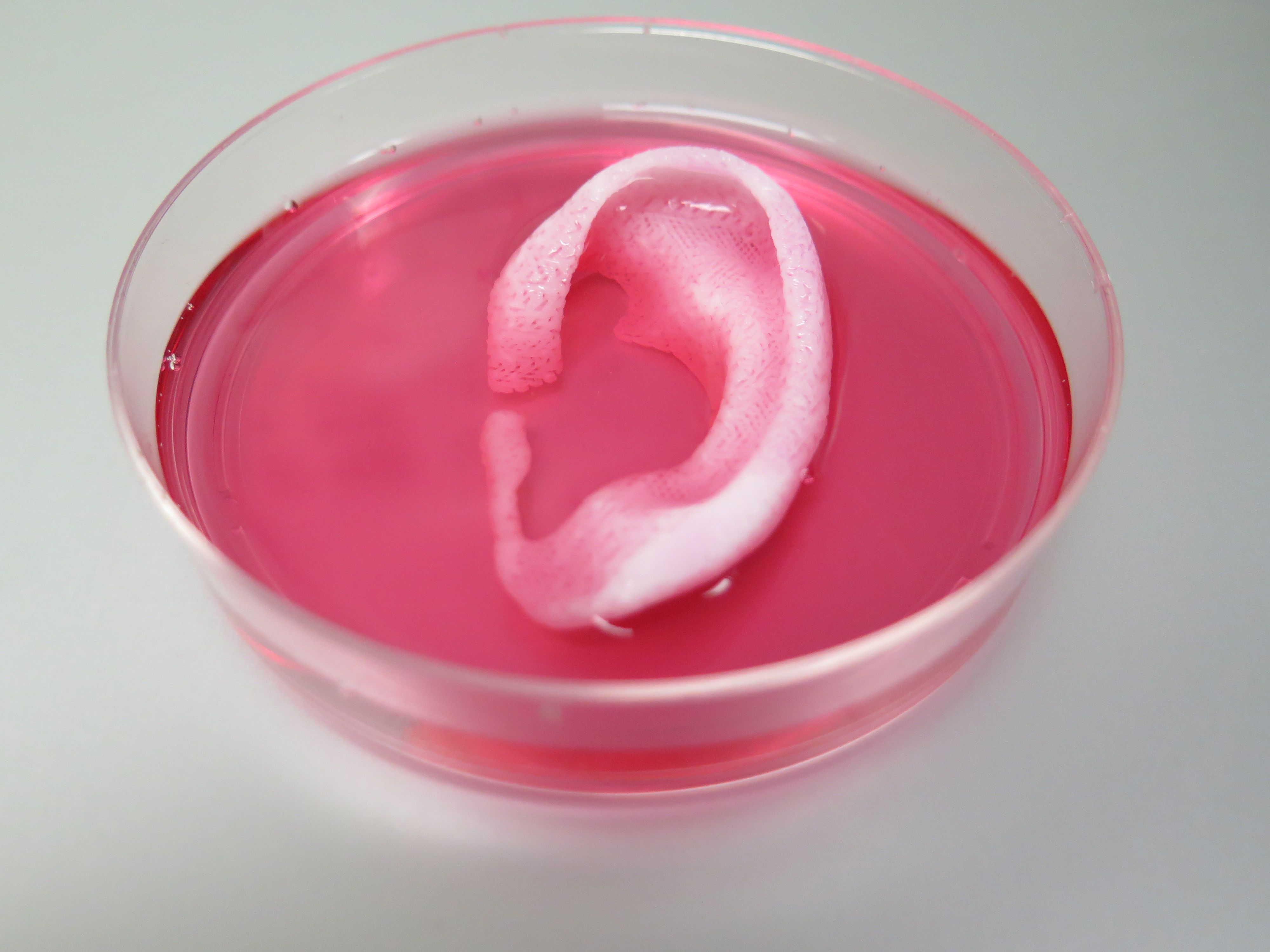
Completed ear and jaw bone structures printed with the Integrated Tissue-Organ Printing System (credit: Wake Forest Baptist Medical Center)

Using a sophisticated, custom-designed 3D printer, regenerative medicine scientists at Wake Forest Baptist Medical Center have proved that it is feasible to print living tissue structures to replace injured or diseased tissue in patients.

Reporting in Nature Biotechnology, the scientists said they printed ear, bone and muscle structures. When implanted in animals, the structures matured into functional tissue and developed a system of blood vessels. Most importantly, these early results indicate that the structures have the right size, strength and function for use in humans.

“This novel tissue and organ printer is an important advance in our quest to make replacement tissue for patients,” said Anthony Atala, M.D., director of the Wake Forest Institute for Regenerative Medicine (WFIRM) and senior author on the study. “It can fabricate stable, human-scale tissue of any shape. With further development, this technology could potentially be used to print living tissue and organ structures for surgical implantation.”

Read more

What happens when a patient is in X-Ray and the X-Ray machine blows up like a bomb killing the patient and staff in the room? Well, a new report shows that it can happen where machines are connected to any network (including the net) because a team of hackers showed in their report how they were they hacked several D.C. and Maryland Hospitals medical devices and numerous machines including life support, X-Rays, etc.


Area hospitals are riddled with cybersecurity flaws that could allow attackers to hack into medical devices and kill patients, a team of Baltimore-based researchers has concluded after a two-year investigation.

Hackers at Independent Security Evaluators say they broke into one hospital’s systems remotely to take control of several patient monitors, which would let an attacker disable alarms or display false information.

The team says it strolled into one hospital’s lobby and used an easily accessible kiosk to commandeer computer systems that track medicine delivery and bloodwork requests — more opportunities for malicious hackers to create mayhem.