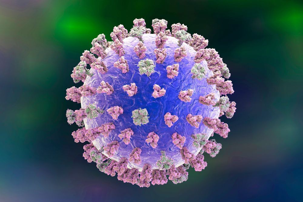
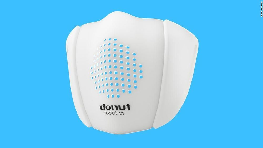
Toggle light / dark theme

Huge US government study to offer genetic counselling

But to generate the kind of long-term data set necessary for breakthroughs in precision medicine — which uses genomic, physiological and other data to tailor treatments to individuals — All of Us must retain these participants, ideally throughout their lives. That’s where genetic counselling comes in.


A firm hired by the National Institutes of Health will work with participants in a research programme that plans to sequence one million genomes.

Revealing the molecular engine that drives pancreatic cancer provides ways to turn it off

Researchers at Georgetown Lombardi Comprehensive Cancer Center have decoded a chain of molecules that are critical for the growth and survival of pancreatic ductal adenocarcinoma (PDAC)—the most common and also the most lethal form of pancreatic cancer.

They say their findings, published in Developmental Cell, suggest that inhibiting this “Yap” biological network may effectively regress early stage PDAC and could be paired with other drugs to halt more advanced stage tumors. Yap inhibitors have been developed and are moving into .

Their study builds upon Georgetown Lombardi research that previously identified Yap as an oncogene central to the initiation of PDAC as well as a variety of other cancers. In the current study employing advanced animal models, they have managed to switch off Yap in pre-established PDAC tumors, and discovered that suppressing Yap blocks the metabolic pathways that provide the fuel and building materials for maintaining the growth of the cancer.

Self-assembled membrane with water-continuous transport pathways for precise nanofiltration

Self-assembled materials are attractive for next-generation materials, but their potential to assemble at the nanoscale and form nanostructures (cylinders, lamellae etc.) remains challenging. In a recent report, Xundu Feng and colleagues at the interdisciplinary departments of chemical and environmental engineering, biomolecular engineering, chemistry and the center for advanced low-dimension materials in the U.S., France, Japan and China, proposed and demonstrated a new approach to prevent the existing challenges. In the study, they explored size-selective transport in the water-continuous medium of a nanostructured polymer template formed using a self-assembled lyotropic H1 (hexagonal cylindrical shaped) mesophase (a state of matter between liquid and solid). They optimized the mesophase composition to facilitate high-fidelity retention of the H1 structure on photoinduced crosslinking.

The resulting nanostructured polymer material was mechanically robust with internally and externally crosslinked nanofibrils surrounded by a continuous aqueous medium. The research team fabricated a with size selectivity at the 1 to 2 nm length scale and water permeabilities of ~10 liters m−2 hour−1 bar−1 μm. The membranes displayed excellent anti-microbial properties for practical use. The results are now published on Science Advances and represent a breakthrough for the potential use of self-assembled membrane-based nanofiltration in practical applications of water purification.

Membrane separation for filtration is widely used in diverse technical applications, including seawater desalination, gas separation, food processing, fuel cells and the emerging fields of sustainable power generation and distillation. During nanofiltration, dissolved or suspended solutes ranging from 1 to 10 nm in size can be removed. New nanofiltration membranes are of particular interest for low-cost treatment of wastewaters to remove organic contaminants including pesticides and metabolites of pharmaceutical drugs. State-of-the-art membranes presently suffer from a trade-off between permeability and selectivity where increased permeability can result in decreased selectivity and vice-versa. Since the trade-off originated from the intrinsic structural limits of conventional membranes, materials scientists have incorporated self-assembled materials as an attractive solution to realize highly selective separation without compromising permeability.

New MRI technique captures image of a brain thinking

An international team of researchers with partial support from the National Institute of Biomedical Imaging and Bioengineering (NIBIB) developed a new MRI technique that can capture an image of a brain thinking by measuring changes in tissue stiffness. The results show that brain function can be tracked on a time scale of 100 milliseconds – 60 times faster than previous methods. The technique could shed new light on altered neuronal activity in brain diseases.

The human brain responds almost immediately to stimuli, but non-invasive imaging techniques haven’t been able to keep pace with the brain. Currently, several non-invasive brain imaging methods measure brain function, but they all have limitations. Most commonly, clinicians and researchers use functional magnetic resonance imaging (fMRI) to measure brain activity via fluctuations in blood oxygen levels. However, a lot of vital brain activity information is lost using fMRI because blood oxygen levels take about six seconds to respond to a stimulus.

Since the mid-1990s, researchers have been able to generate maps of tissue stiffness using an MRI scanner, with a non-invasive technique called magnetic resonance elastography (MRE). Tissue stiffness can’t be measured directly, so instead researchers use MRE to measure the speed at which mechanical vibrations travel through tissue. Vibrations move faster through stiffer tissues, while vibrations travel through softer tissue more slowly; therefore, tissue stiffness can be determined. MRE is most commonly used to detect the hardening of liver tissue but has more recently been applied to other tissues like the brain.

Rapid tissue regeneration induced by intracellular ATP delivery—A preliminary mechanistic study

Circa 2017


We have reported a new phenomenon in acute wound healing following the use of intracellular ATP delivery—extremely rapid tissue regeneration, which starts less than 24 h after surgery, and is accompanied by massive macrophage trafficking, in situ proliferation, and direct collagen production. This unusual process bypasses the formation of the traditional provisional extracellular matrix and significantly shortens the wound healing process. Although macrophages/monocytes are known to play a critical role in the initiation and progression of wound healing, their in situ proliferation and direct collagen production in wound healing have never been reported previously. We have explored these two very specific pathways during wound healing, while excluding confounding factors in the in vivo environment by analyzing wound samples and performing in vitro studies. The use of immunohistochemical studies enabled the detection of in situ macrophage proliferation in ATP-vesicle treated wounds. Primary human macrophages and Raw 264.7 cells were used for an in vitro study involving treatment with ATP vesicles, free Mg-ATP alone, lipid vesicles alone, Regranex, or culture medium. Collagen type 1α 1, MCP-1, IL-6, and IL-10 levels were determined by ELISA of the culture supernatant. The intracellular collagen type 1α1 localization was determined with immunocytochemistry. ATP-vesicle treated wounds showed high immunoreactivity towards BrdU and PCNA antigens, indicating in situ proliferation. Most of the cultured macrophages treated with ATP-vesicles maintained their classic phenotype and expressed high levels of collagen type 1α1 for a longer duration than was observed with cells treated with Regranex. These studies provide the first clear evidence of in situ macrophage proliferation and direct collagen production during wound healing. These findings provide part of the explanation for the extremely rapid tissue regeneration, and this treatment may hold promise for acute and chronic wound care.

Wound healing is a complex and dynamic process involving the replacement of devitalized and missing structures. The traditional view of wound healing is that it involves hemostasis, inflammation, proliferation, and remodeling, and these steps result in a lag of 3–6 d before reepithelialization starts [1,2]. We have discovered that the intracellular delivery of adenosine triphosphate using ATP-vesicles as an acute wound treatment enhances wound healing [3,4]. The most unprecedented finding was that new tissue started to generate within 24 h, and it continued to grow to eliminate the wound cavity quickly [4–6]. This growth was attained by early and massive monocyte/macrophage trafficking, proliferation, and fast collagen production for direct formation of extracellular matrix (ECM).

Nothing found for Boosting Nad Improves Age%25E2%2580%2590Related Hearing Impairment In Mice

Today, we want to spotlight a recent study showing that boosting nicotinamide adenine dinucleotide (NAD+) levels in mice prevents age-related hearing loss.

What is β-Lapachone?

β-Lapachone is a quinone-containing compound that was originally isolated from the lapacho tree in South America. It is worth noting that this tree has been used as a herbal medicine for a number of South and Central American indigenous peoples and that the bark of the tree is sometimes used for making a herbal tea called taheebo.

/* */