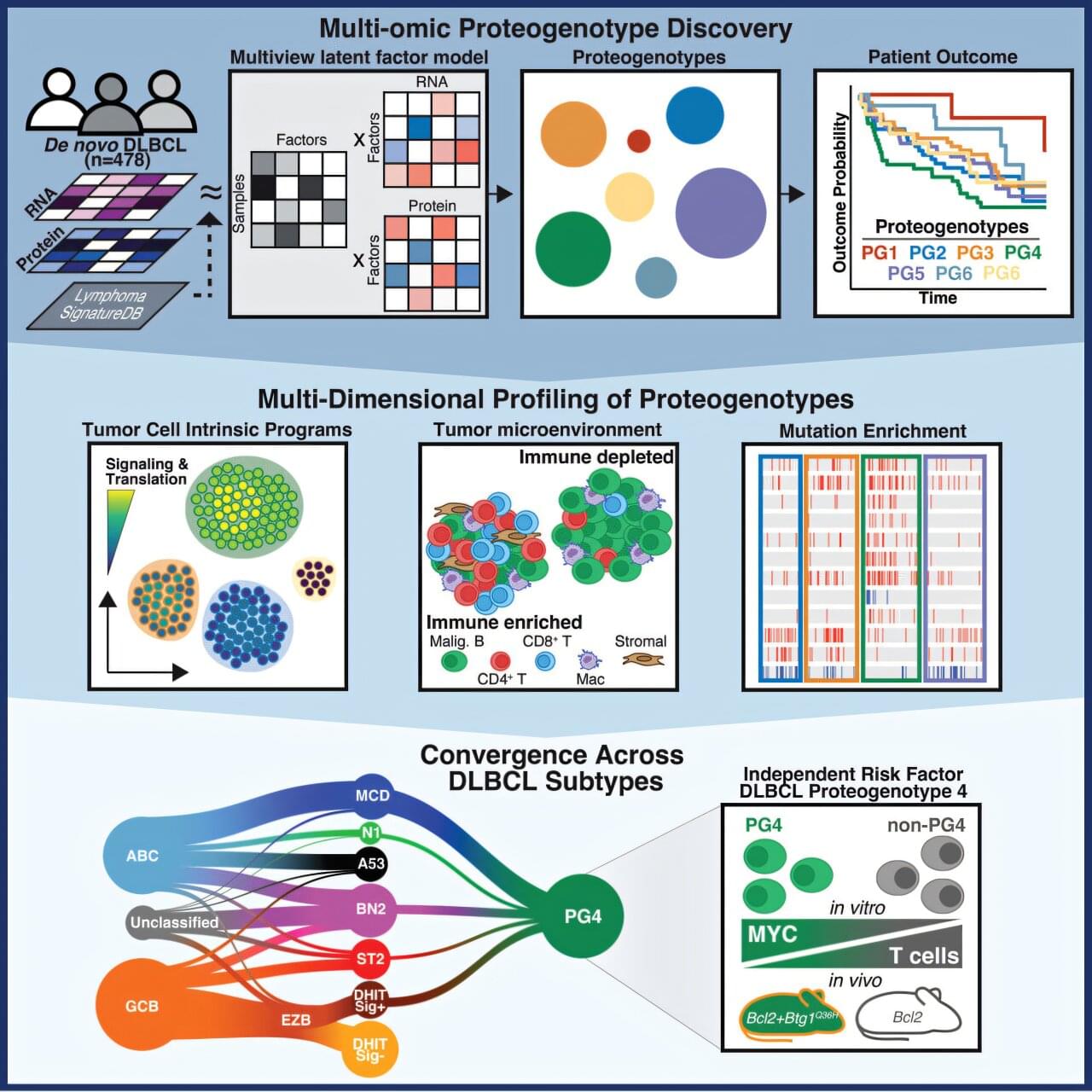
Researchers led by Universitätsmedizin Frankfurt and Goethe University Frankfurt have identified how particularly aggressive forms of lymphoma can be recognized. By combining genetic and proteomic analyses, the scientists identified biological characteristics of tumors, particularly in high-risk patients for whom standard therapy offers little chance of cure. In the future, such patients could receive alternative, more effective therapies directly. In addition, experimental laboratory research provided initial clues to potential therapeutic targets. The study is published in Cancer Cell.
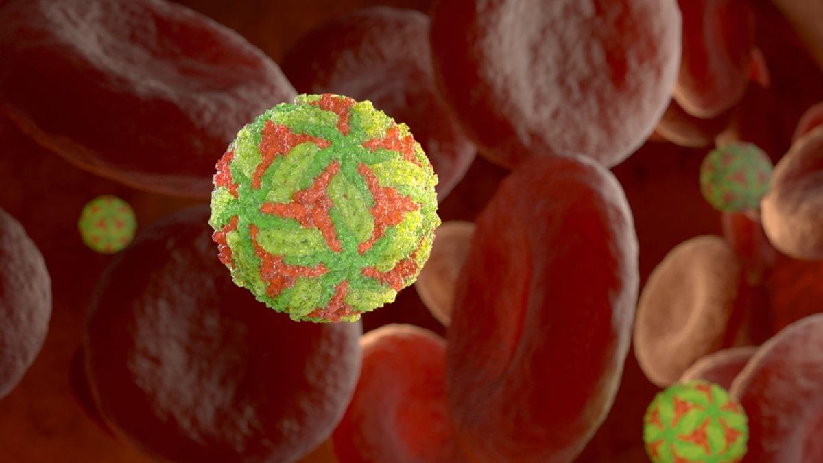
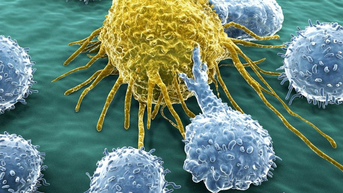
With more than 150,000 new cases worldwide each year, diffuse large B-cell lymphoma (DLBCL) is the most common aggressive form of lymphoma. Following diagnosis, patients typically receive a standard treatment regimen consisting of a therapeutic antibody and chemotherapy (R-CHOP or Pola-R-CHP), and nearly two-thirds of patients have a good chance of being cured. However, more than one-third of patients experience a relapse after treatment, or their tumors fail to respond to therapy, requiring alternative treatments such as CAR T-cell therapy.
The varying effectiveness of standard therapy is due to the considerable molecular heterogeneity of the disease. Researchers have therefore long been searching for molecular tumor characteristics that would allow them to distinguish among different DLBCL subtypes and treat them more specifically.