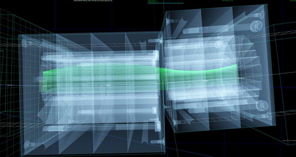
Toggle light / dark theme

The magnetization-prepared rapid gradient-echo (MP-RAGE) T1-weighted high resolution structural MRI is a mainstay tool used to identify morphometric biomarkers of disease conditions, progression and treatment effects despite a critical limitation: the relaxation signal on which inferences are based is nearly indistinguishable for gray matter vs. blood flow (Lu et al., 2004; Wright et al., 2008). Thus, apparent reported morphometric findings might be at least partially related to transient changes in blood flow or other physiological signals.

Consistent with this technical limitation, using a standard analysis technique, voxel based morphometry (VBM), we recently reported that a single dose of a medication had “apparent” effects on T1-weighted MRIs (Franklin et al., 2013). Specifically, we observed medication-induced decreases in gray matter volume in the anterior cingulate and other regions that overlapped with changes in brain blood flow (perfusion). Similarly, others have shown effects of medication on T1-weighted scans that are likely transient. For example, acute levodopa administration altered gray matter indices on T1-weighted images in the midbrain (Salgado-Pineda et al., 2006). Further, in a well-controlled longitudinal VBM study of patients with attention deficit hyperactivity disorder (ADHD), Hoekzema et al.

Objectives: Proximal median nerve (PMN) neuropathies are caused by lesions proximal to the carpal tunnel, which include the forearm, elbow, upper arm, and brachial plexus. Differentiating between carpal tunnel syndrome and PMN neuropathies is important to guide management and is based on clinical, electrodiagnostic (EDX), and ultrasound (US) findings. This study describes the clinical, EDX, and US features in 62 patients with PMNs.

Methods: All patients underwent EDX studies, and 52 (83.9%) had a US study. The patients were assigned to one of the following four localization zones of PMN neuropathies based on clinical and EDX criteria: Zone 1: extends from the fascicles in the brachial plexus contributing to the median nerve to the innervation of the pronator teres (PT); Zone 2: distal to the branch to the PT and proximal to the origin of the anterior interosseous nerve (AIN); Zone 3: involves the origin of the AIN; and Zone 4: distal to the origin of the AIN and proximal to the carpal tunnel. The localization was based on the pattern of muscle weakness, topography of EMG abnormalities, and US study findings.

Results: The anatomical locations of the PMN neuropathies based on clinical, EDX, and US findings were as follows: Zone 1 in 38 patients (61.3%), Zone 2 in 6 patients (9.7%), Zone 3 in 7 patients (11.3%), and Zone 4 in 11 patients (17.7%). The most common etiology among all 62 patients was iatrogenic injury (30 [48.4%]), followed by non-iatrogenic trauma (20 [32.2%]). The following EDX findings were noted: prolonged distal motor latency (29 [46.8%]), decreased motor nerve conduction velocity in the forearm (22 [35.5%]), low amplitude or absent compound muscle action potentials (50 [80.6%]), and abnormal or absent sensory nerve action potentials (50 [80.6%]). Of the 52 (83.9%) patients who underwent US studies, a total of 22 (42.3%) patients showed an increased cross-sectional area of the median nerve.

Scientists at Northwestern University and University of California San Diego have developed a new, potent injectable therapy that can protect the heart from damage after a heart attack.

The therapeutic approach comprises specially designed polymers that act like proteins. These protein-like polymers (PLPs) “grab” onto regulatory proteins, which blunt the body’s natural healing process, in heart tissue. With those proteins out of the way, the healing proteins are free to do their job — preventing stress and inflammation.


Protein-like polymer demonstrated improved heart health in animal experiments.

Heart attacks cause long-term damage that ultimately leads to heart failure. New treatment protects the heart from long-term damage after a heart attack.

For decades, researchers have been exploring ways to harness the power of the immune system to treat cancer. One breakthrough is cell therapy, often called ‘living drugs.’ This is a form of immunotherapy that uses immune cells from a patient or a healthy donor. With advanced engineering techniques, scientists enhance these cells to recognize better and attack cancer.

“During the late 1980s and 1990s, cancer researchers started exploring ways to advance immunotherapy by transferring immune cells into a patient to attack cancer cells,” says stem cell transplant and cellular therapy specialist Hind Rafei, M.D. “They recognized that immune cells found inside tumors could help destroy cancer cells, leading to the development of one of the earliest forms of cell therapy — tumor-infiltrating lymphocytes (TILs).”


Cell therapy is a form of immunotherapy that uses immune cells from a patient or a healthy donor to treat cancer. Learn about the types of cell therapy from stem cell transplant and cellular therapy specialist Hind Rafei, M.D.