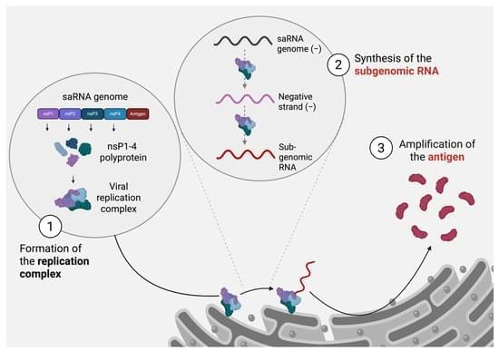
Toggle light / dark theme

Microscale hydrogel fibers could enable imaging inside tiny tissue structures

Researchers have developed light-transmitting hydrogel fibers that are just hundreds of micrometers in diameter. With further development, these soft fibers could one day make it possible to use imaging techniques to detect early breast cancer hidden inside very small breast ducts.

“While traditional, relatively rigid fiber probes may cause mechanical damage when entering narrow, curved or soft tissue spaces, our fibers are very soft with mechanical properties more similar to those of human soft tissues,” said research team leader Yu Zhang from Harbin Engineering University in China. “We made these fibers using a draw-spinning method that was inspired by spider-silk spinning.”

In research appearing in Optics Express, the researchers describe how they tested the new hydrogel fibers by incorporating them into an imaging system and using it to analyze standard pathology-stained breast tissue sections. The imaging system successfully reconstructed the microscopic features used by pathologists to evaluate tumors and, when combined with artificial intelligence algorithms, distinguished tumor subtypes with an accuracy of 93.97%.

Ultra-precise technology can count damaged DNA fragments

The Korea Research Institute of Standards and Science has developed an ultrasensitive immunoassay-based analytical platform that can detect and quantify trace amounts of “Small Excised Damaged DNA (sedDNA)” fragments generated during cellular DNA repair. This technology enables highly sensitive detection with quantification down to the level of several thousand molecules, measuring up to 22 times more DNA fragments than conventional methods. It provides a new analytical foundation for comparing DNA repair capacity between individuals and studying cellular responses to anticancer drugs and carcinogenic agents.

Human DNA is continuously exposed to damage from ultraviolet light, chemical agents, smoking and normal metabolic processes. If such damage is not properly repaired, mutations can accumulate and lead to aging and diseases such as cancer. To maintain genomic stability, cells activate the Nucleotide Excision Repair (NER) system, which removes damaged DNA segments and replaces them with newly synthesized DNA. The small excised DNA fragments generated during this process serve as important indicators of DNA repair efficiency and kinetics, providing a valuable tool for studying disease mechanisms and predicting treatment responses.

Copper-induced cell death stimulates antitumor immune cell responses

A preclinical study from researchers at The University of Texas MD Anderson Cancer Center, published today in Cell, details a connection between the immune system and cuproptosis, a type of copper-induced cell death. The findings suggest a new approach to help overcome resistance to immunotherapy.

The study, led by Boyi Gan, Ph.D., professor of Experimental Radiation Oncology, demonstrates that cancer cells undergoing cuproptosis release signals that activate the immune system. Significantly, this study is among the first to demonstrate that cuproptosis can actively engage the immune system and enhance responses to immunotherapy. In preclinical models, a combination approach of cuproptosis-inducing treatment along with anti-PD-L1 immunotherapy significantly slowed tumor growth.

“This study reveals a previously unrecognized partnership between the immune system and cuproptosis,” Gan said. “Importantly, because the cuproptosis-inducing agents used in our studies already have clinical experience and favorable safety profiles, these findings may offer a practical path toward developing new combination treatments for patients whose cancers no longer respond to immunotherapy.”

The Revolving Door of Adenovirus Cell Entry: Not All Pathways Are Equal

An interesting review on adenoviral cell entry and trafficking. Its discussion of how species B adenoviruses tolerate lower endosomal pH and accumulate in later-endosomal compartments before escaping were particularly intriguing. Link.


Adenoviruses represent exceptional candidates for wide-ranging therapeutic applications, from vectors for gene therapy to oncolytics for cancer treatments. The first ever commercial gene therapy medicine was based on a recombinant adenovirus vector, while most recently, adenoviral vectors have proven critical as vaccine platforms in effectively controlling the global coronavirus pandemic. Here, we discuss factors involved in adenovirus cell binding, entry, and trafficking; how they influence efficiency of adenovirus-based vectors; and how they can be manipulated to enhance efficacy of genetically modified adenoviral variants. We focus particularly on endocytosis and how different adenovirus serotypes employ different endocytic pathways to gain cell entry, and thus, have different intracellular trafficking pathways that subsequently trigger different host antiviral responses.

Opposing protein pathways steer skin stem cells toward renewal or repair

Two proteins with opposing functions orchestrate the development and maintenance of healthy skin, Stanford Medicine researchers have found. Modulating their activity with topical drugs could reduce inflammation, aid wound healing and slow or halt the growth of skin cancer, the researchers believe. The findings are published in the journal Science.

The proteins are part of a family called ubiquitin-like proteins. Ubiquitination controls the targeted destruction and disposal of unneeded proteins in a cell. But in the skin, certain ubiquitin-like proteins instead switch on or off wide swaths of genes involved in cellular growth and development, the study found. In particular, they trigger progenitor (stem) cells in the lower layer of the skin to either mature and migrate to the skin surface or to self-renew.

“These two ubiquitin-like protein systems are remarkably dedicated and opposite in their functions,” said Paul Khavari, MD, Ph.D., chair of dermatology at the Stanford School of Medicine and senior author of the study. “One promotes the stem-cell state while the other drives differentiation. It’s like having two opposing forces that determine a cell’s fate.”

Iron accumulation in the brain may contribute to neurodegeneration

Neurodegenerative diseases affect tens of millions of people worldwide. Among these, Alzheimer’s and Parkinson’s diseases are the most common; in the United States alone, the Alzheimer’s Disease Association and Parkinson’s Foundation report roughly 7 million people with Alzheimer’s and another million with Parkinson’s. An intriguing clue lies in the tangled mystery of neurodegeneration that scientists are working to solve: iron accumulation.

Scientists have noticed that iron can slowly build up inside neurons. Early in life, this iron accumulation appears to have little effect on neuronal function. However, later in life, it can contribute to a slow neuronal demise. Salk Institute researchers studied nerve cells to figure out whether and how this iron accumulation relates to neurodegenerative diseases. They found that the excess iron stuck in neurons lowers the cells’ defenses, making them more vulnerable to stressors and other cellular insults through a process they named chronoferroptosis.

The study, published in Cell Death Discovery on June 18, 2026, points to iron accumulation as a key target in the effort to predict, prevent and treat neurodegenerative diseases.

First 3D views of human cone opsins reveal how daylight vision reacts so fast

The retina of the human eye contains 6–7 million cone cells. These cells contain light-sensitive proteins known as cone opsins. They enable us to perceive our surroundings in detail in daylight. They allow us to see the world in thousands of colors: red strawberries, green leaves, the blue sky. They also enable us to see all the objects around us clearly. And they allow us to perceive fast movements, such as the rush of a train or the flight of a dragonfly.

Often, however, these all-rounders of daylight vision are also involved in retinal diseases. Impairment of cone receptor function, caused by genetic mutations or other degenerative processes, can lead to disorders such as color blindness and age-related macular degeneration (AMD), a disease affecting the central retina and causing progressive vision loss.

In a new study, Polina Isaikina and Sarah L. Schmidt, two researchers from the Center for Life Sciences at PSI, have succeeded for the first time in determining the three-dimensional structure of human cone opsins in their dark state and showing how their molecular architecture enables their rapid activation by light.

/* */