Gould’s thesis has sparked widespread debate ever since, with some advocating for determinism and others supporting contingency. In his 1952 short story A Sound of Thunder, science fiction author Ray Bradbury recounted how a time traveler’s simple act of stepping on a butterfly in the age of the dinosaurs changed the course of the future. Gould made a similar point: “Alter any early event, ever so slightly and without apparent importance at the time, and evolution cascades into a radically different channel.”
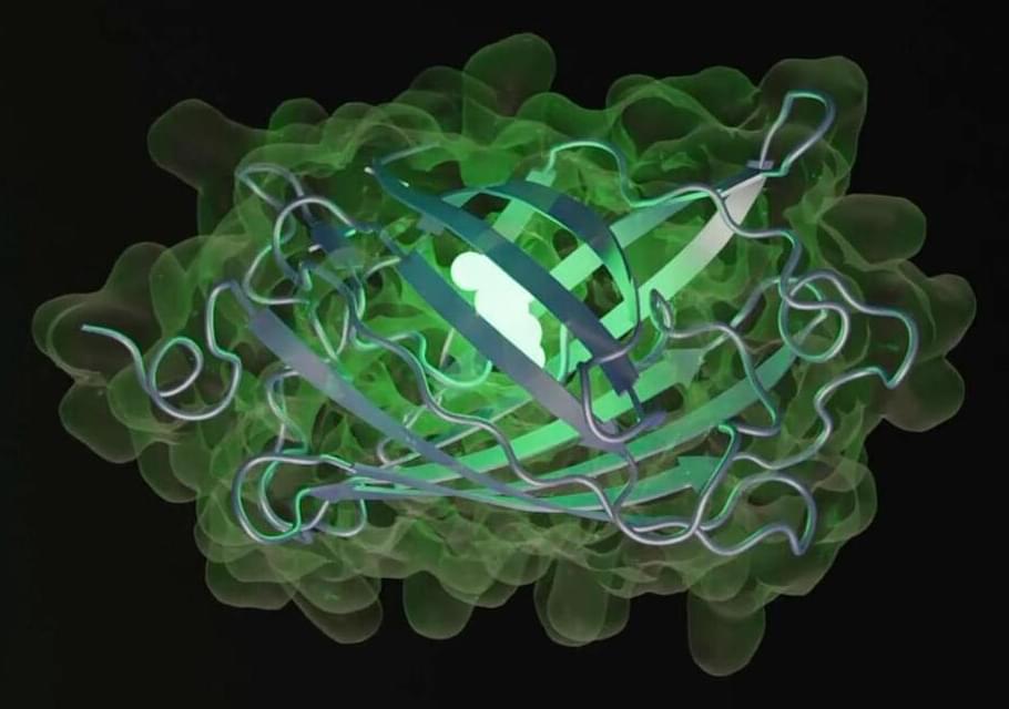
Scientists have been exploring this problem through experiments designed to recreate evolution in the lab or in nature, or by comparing species that have emerged under similar conditions. Today, a new avenue has opened up: AI. In New York, a group of former researchers from Meta — the parent company of social networks Facebook, Instagram, and WhatsApp — founded EvolutionaryScale, an AI startup focused on biology. The EvolutionaryScale Model 3 (ESM3) system created by the company is a generative language model — the same kind of platform that powers ChatGPT. However, while ChatGPT generates text, ESM3 generates proteins, the fundamental building blocks of life.
ESM3 feeds on sequence, structure, and function data from existing proteins to learn the biological language of these molecules and create new ones. Its creators have trained it with 771 billion data packets derived from 3.15 billion sequences, 236 million structures, and 539 million functional traits. This adds up to more than one trillion teraflops (a measure of computational performance) — the most computing power ever used in biology, according to the company.