Toggle light / dark theme

Ideally, the nanopore dimensions should be comparable to those of the analyte for the presence of the analyte to produce a measurable change in the ionic current amplitude above the noise level. Nanopores can be formed in several ways, with a wide range of pore diameters. Biological nanopores are formed by the self-assembly of either protein subunits, peptides or even DNA scaffolds in lipid bilayers or block copolymer membranes1,3,6,17,18. They possess atomically precise dimensions controlled by biopolymer sequences, providing the ability to recognize biomolecules with constriction diameters of ~1–10 nm. Solid-state nanopores are crafted in thin inorganic or plastic membranes (for example, SiNx), which allows the nanopores to have extended diameters of up to hundreds of nanometres, permitting the entry or analysis of large biomolecules and complexes. The tools for fabricating solid-state nanopores, which include electron/ion milling4,5, laser-based optical etching19,20 and the dielectric breakdown of ultrathin solid membranes21,22, can be used to manipulate nanopore size at the nanometre scale, but allow only limited control over the surface structure at the atomic level in contrast to biological nanopores. The chemical modification and genetic engineering of biological nanopores, or the introduction of biomolecules to functionalize solid-state nanopores23, can further enhance the interactions between a nanopore and analytes, improving the overall sensitivity and selectivity of the device2,17,24,25,26. This feature allows nanopores to controllably capture, identify and transport a wide variety of molecules and ions from bulk solution.

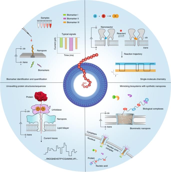
Nanopore technology was initially developed for the practicable stochastic sensing of ions and small molecules2,27,28. Subsequently, many developmental efforts were focused on DNA sequencing1,7,8,9. Now, however, nanopore applications extend well beyond sequencing, as the methodology has been adapted to analyse molecular heterogeneities and stochastic processes in many different biochemical systems (Fig. 1). First, a key advantage of nanopores lies in their ability to successively capture many single molecules one after the other at a relatively high rate, which allows nanopores to explore large populations of molecules at the single-molecule level in reasonable timeframes. Second, nanopores essentially convert the structural and chemical properties of the analytes into a measurable ionic current signal, even achieving enantiomer discrimination29. The technology can be used to report on multiple molecular features while circumventing the need for labelling chemistries, which may complicate the overall analysis process and affect the molecular structures. For example, nanopores can discriminate nearly 13 different amino acids in a label-free manner, including some with minute structural differences30. An important aspect is the ability of nanopores to identify species31 that lack suitable labels for signal amplification or whose information is hidden in the noise of analytical devices. Consequently, nanopores may serve well in molecular diagnostic applications required for precision medicine, which achieves the identification of nucleic acid, protein or metabolite analytes and other biomarkers11,32,33,34,35. Third, nanopores provide a well-defined scaffold for controllably designing and constructing biomimetic systems, which involve a complex network of biomolecular interactions. These nanopore systems track the binding dynamics of transported biomolecules as they interact with nanopore surfaces, hence serving as a platform for unravelling complex biological processes (for example, the transport properties of nuclear pore complexes)36,37,38,39. Fourth, chemical groups can be spatially aligned within a protein nanopore, providing a confined chemical environment for site-selective or regioselective covalent chemistry. This strategy has been used to engineer protein nanoreactors to monitor bond-breaking and bond-making events40,41.

Here we discuss the latest advances in nanopore technologies beyond DNA sequencing and the future trajectory of the field, as well as the opportunities and main challenges for the next decade. We specifically address the emerging nanopore methods for protein analysis and protein sequencing, single-molecule covalent chemistry, single-molecule analysis of clinical samples and insights into the use of biomimetic pores for analysing complex biological processes.
Circa 2017 face_with_colon_three


LA JOLLA—Salk scientists have created a new version of the CRISPR/Cas9 genome editing technology that allows them to activate genes without creating breaks in the DNA, potentially circumventing a major hurdle to using gene editing technologies to treat human diseases.

Most CRISPR/Cas9 systems work by creating “double-strand breaks” (DSBs) in regions of the genome targeted for editing or for deletion, but many researchers are opposed to creating such breaks in the DNA of living humans. As a proof of concept, the Salk group used their new approach to treat several diseases, including diabetes, acute kidney disease, and muscular dystrophy, in mouse models.

In a clinical trial, the first patient has received a single dose of a new human immunodeficiency virus (HIV) gene editing therapy, researchers at the Lewis Katz School of Medicine at Temple University and Excision BioTherapeutics, Inc have reported.

In a collaborative effort, the researchers are currently running a phase 1/2 clinical trial to evaluate the safety and efficacy of their therapy, called EBT-101, which is based on gene editing technology known as CRISPR.

“Nearly 40 million people worldwide suffer from the effects of HIV, and more than 40 years after the discovery of HIV/AIDS, there still are no curative treatments,” said Professor Kamel Khalili, who helped lead the trial, in a statement.

When the inside of a mollusk shell shimmers in sunlight, the iridescence isn’t produced by colored pigments but by tiny physical structures self-assembled from living cells and inorganic components. Now, a team of researchers at the Department of Energy’s Lawrence Berkeley National Laboratory (Berkeley Lab) has developed a platform to mimic this self-assembly ability by engineering living cells to act as a starting point for building composite materials.

Engineered living (ELMs) use living as “materials scaffolds” and are a new class of material that might open the door to self-healing materials and other advanced applications in bioelectronics, biosensing, and smart materials. Such materials could mimic emergent properties found in nature—where a complex system has properties that the individual components do not have—such as iridescence or strength.

Borrowing from this complexity seen in nature, the Berkeley Lab researchers engineered a bacterium that can attach a wide range of nanomaterials to its cell surface. They can also precisely control the makeup and how densely packed the components are, creating a stable hybrid living material. The study describing their work was recently published in ACS Synthetic Biology.

Chromosome-level engineering is a completely different beast: it’s like rearranging multiple paragraphs or shifting complete sections of an article and simultaneously hoping the changes add capabilities that can be passed onto the next generation.

Reprogramming life isn’t easy. Xiao Zhu’s DNA makeup is built from genetic letters already optimized by eons of evolutionary pressure. It’s no surprise that tinkering with an established genomic book often results in life that’s not viable. So far, only yeast have survived the rejiggering of their chromosomes.

The new study, published in Science, made the technology possible for mice. The team artificially fused together chunks from mice chromosomes. One fused pair made from chromosomes four and five was able to support embryos that developed into healthy—if somewhat strangely behaved—mice. Remarkably, even with this tectonic shift to their normal genetics, the mice could reproduce and pass on their engineered genetic quirks to a second generation of offspring.

Small but mighty, lysosomes play a surprisingly important role in cells despite their diminutive size. Making up only 1–3% of the cell by volume, these small sacs are the cell’s recycling centers, home to enzymes that break down unneeded molecules into small pieces that can then be reassembled to form new ones. Lysosomal dysfunction can lead to a variety of neurodegenerative or other diseases, but without ways to better study the inner contents of lysosomes, the exact molecules involved in diseases—and therefore new drugs to target them—remain elusive.

A new method, reported in Nature on Sept. 21, allows scientists to determine all the molecules present in the lysosomes of any cell in mice. Studying the contents of these molecular recycling centers could help researchers learn how the improper degradation of cellular materials leads to certain diseases. Led by Stanford University’s Monther Abu-Remaileh, institute scholar at Sarafan ChEM-H, the study’s team also learned more about the cause for a currently untreatable neurodegenerative known as Batten disease, information that could lead to new therapies.

“Lysosomes are fascinating both fundamentally and clinically: they supply the rest of the cell with nutrients, but we don’t always know how and when they supply them, and they are the places where many diseases, especially those that affect the brain, start,” said Abu-Remaileh, who is an assistant professor of chemical engineering and of genetics.

For the memory prosthetic, the team focused on two specific regions: CA1 and CA3, which form a highly interconnected neural circuit. Decades of work in rodents, primates, and humans have pointed to this neural highway as the crux for encoding memories.

The team members, led by Drs. Dong Song from the University of Southern California and Robert Hampson at Wake Forest School of Medicine, are no strangers to memory prosthetics. With “memory bioengineer” Dr. Theodore Berger—who’s worked on hijacking the CA3-CA1 circuit for memory improvement for over three decades—the dream team had their first success in humans in 2015.

The central idea is simple: replicate the hippocampus’ signals with a digital replace ment. It’s no easy task. Unlike computer circuits, neural circuits are non-linear. This means that signals are often extremely noisy and overlap in time, which bolsters—or inhibits—neural signals. As Berger said at the time: “It’s a chaotic black box.”

The genetic encoding of ncAAs with distinct chemical, biological, and physical properties requires the engineering of bioorthogonal translational machinery, consisting of an evolved aminoacyl-tRNA synthetase/tRNA pair and a “blank” codon. To achieve this, the researchers mimicked the ibis’ ability to synthesize sTyr and incorporate it into proteins.

The Xiao lab employed a mutant amber stop codon to encode the desired sulfotransferase, resulting in a completely autonomous mammalian cell line capable of biosynthesizing sTyr and incorporating it with great precision into proteins.

These engineered cells, the authors wrote, can produce “site-specifically sulfated proteins at a higher yield than cells fed exogenously with the highest level of sTyr reported in the literature.” They used the cells to prepare highly potent thrombin inhibitors with site-specific sulfation.

Can you believe that we have a state-of-the-art laboratory in space?

The International Space Station has been in low Earth orbit since 1998. Astronauts started to use the station in November 2000, when a module that provided a long-term life support and control system was added to the first two modules.

Since then, the International Space Station has hosted more than 250 astronauts from 20 countries, most of which have been from Russia and the US.