G-protein-coupled receptors (GPCRs) are one of the largest families of cell surface proteins in the human body that recognize hormones, neurotransmitters, and drugs. These receptors regulate a wide range of physiological processes and are the targets of more than 30% of currently marketed drugs. The histamine H1 receptor (H1R) is one such GPCR subtype that plays a key role in mediating allergic reactions, inflammation, vascular permeability, airway constriction, wakefulness, and cognitive functions in the human body. While antihistamines primarily target H1R, current drugs can exhibit limited therapeutic efficacy, prompting researchers to look at H1R ligands from new perspectives.
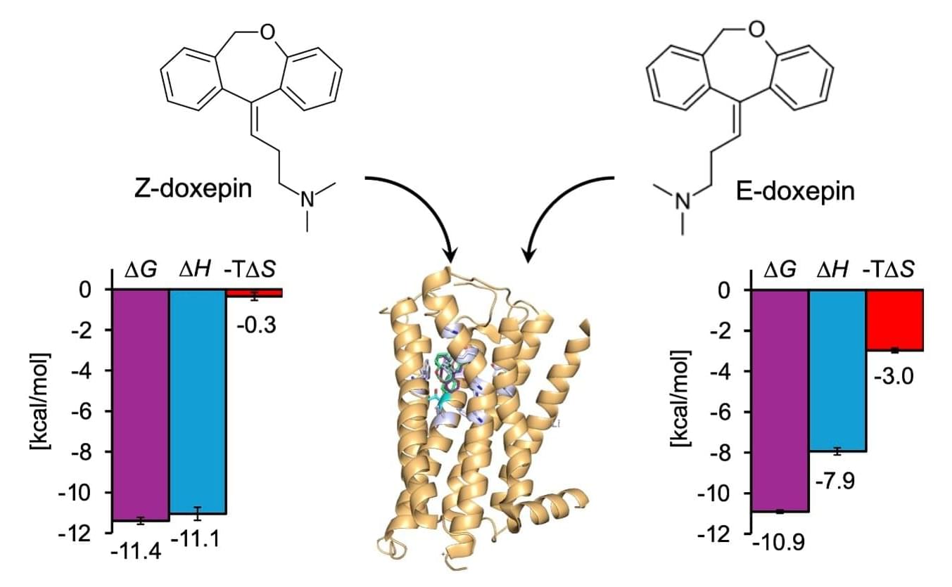
Recently, the importance of drug design based not only on the affinity or binding energy between a compound and its target protein, but also on its components, enthalpy, and entropy, has been recognized as crucial for rational drug design. In particular, enthalpy–entropy compensation has emerged as a key concept for understanding ligand selectivity and isomer specificity. However, direct experimental measurement of these thermodynamic parameters has been limited to cell surface proteins, such as GPCRs.
Addressing this gap, a research team led by Professor Mitsunori Shiroishi from the Department of Life System Engineering, Tokyo University of Science (TUS), Japan, systematically investigated the binding thermodynamics of the H1R. The team included Mr. Hiroto Kaneko (first-year doctoral student) and Associate Professor Tadashi Ando from TUS, among others. Their study was published online in ACS Medicinal Chemistry Letters on January 26, 2026.