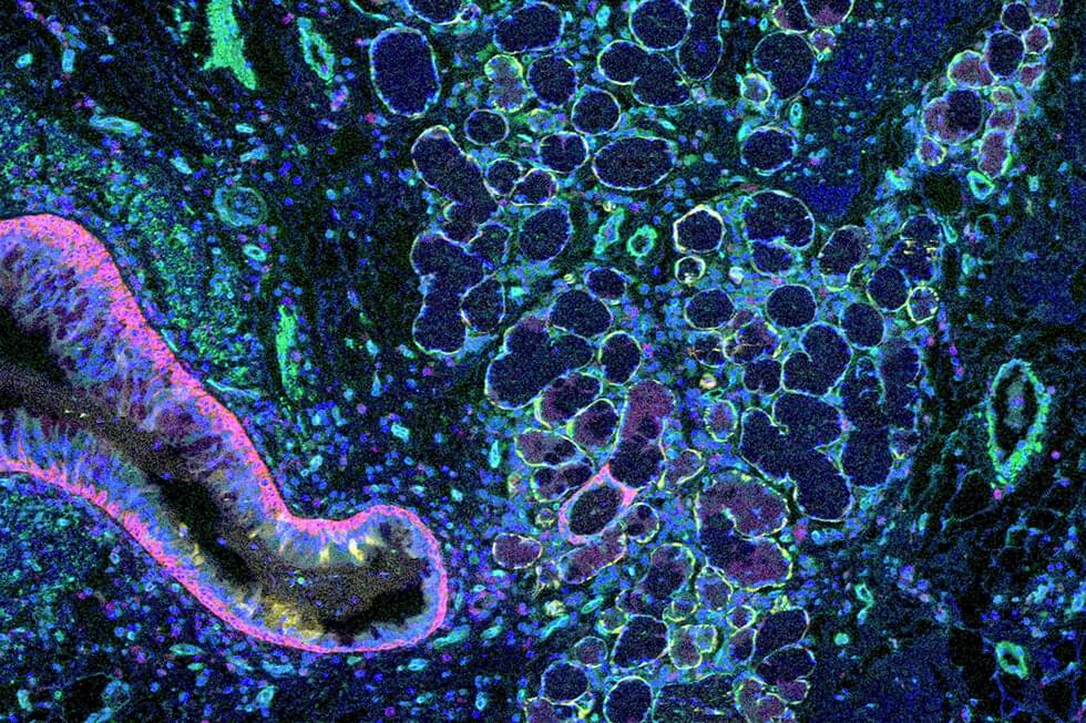
Weill Cornell Medicine researchers have developed a computational method to map the architecture of human tissues in unprecedented detail. Their approach promises to accelerate studies on organ-scale cellular interactions and could enable powerful new diagnostic strategies for a wide range of diseases.
The method, published Oct. 31 in Nature Methods, grew out of the scientists’ frustration with the gap between classical microscopy and modern single-cell molecular analysis. “Looking at tissues under the microscope, you see a bunch of cells that are grouped together spatially—you see that organization in images almost immediately,” said lead author Junbum Kim, a graduate student in physiology and biophysics at Weill Cornell Medicine.
“Now, cell biologists have gained the ability to examine individual cells in tremendous detail, down to which genes each cell is expressing, so they’re focused on the cells instead of focusing on the tissue structure,” he said.


Comments are closed.