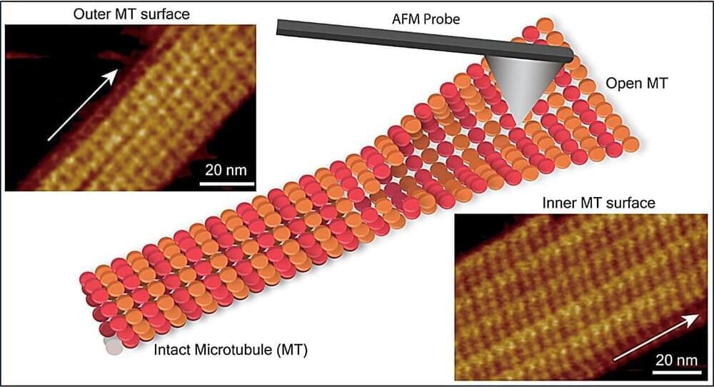
In a study recently published in the journal Nano Letters, researchers from Nano Life Science Institute (WPI-NanoLSI), Kanazawa University, Kanazawa, Japan, used frequency-modulated atomic force microscopy to reveal the submolecular structure of microtubule (MT) inner surface and visualize structural defects in the MT lattice, providing valuable insights into the complex dynamic processes that regulate microtubule function.
Microtubules (MTs), a key component of the cytoskeleton in eukaryotic cells, serve as scaffolds and play vital roles in cellular processes such as cell division, cell migration, intracellular transport, and trafficking. MTs are composed of α-tubulin and β-tubulin proteins, which polymerize into dimers and assemble into linear protofilaments that form a cylindrical lattice.
Traditional methods like X-ray crystallography and cryo–electron microscopy have provided structural insights into MTs but involve complex sample preparation and data analysis. There remains a need for techniques that can examine MT structural features, assembly dynamics, and lattice defects at submolecular resolution under physiological conditions.
